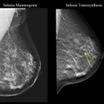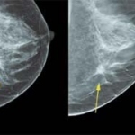
Breast Care Solutions from Siemens at the German Radiology Congress
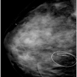
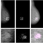
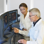
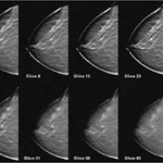
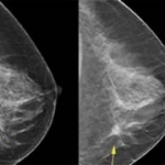
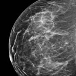
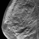
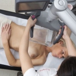
Siemens Healthcare was presenting its latest solutions for the early detection and treatment of breast cancer at the German Radiology Congress in Berlin. These Breast Care Solutions include a variety of imaging procedures, such as ultrasound, mammography, and magnetic resonance imaging (MRI), supplemented by IT and laboratory diagnostic solutions. Siemens places special focus on the third…