Evaluating breast tomosynthesis for the Netherlands
Combining a scientific research laboratory with specialised clinic, the Netherlands Cancer Institute, in the Antoni van Leeuwenhoek Hospital (NKI-AVL), in Amsterdam, aims for a unique interaction of scientific research and clinical application.
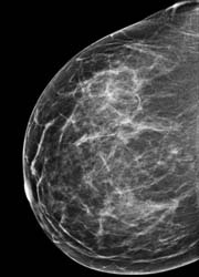
to date using breast tomosynthesis. In two of the 300 cases, tomosynthesis was clearly better. In one case the patient had mammography, ultrasound and a biopsy in another hospital, but came here for a second opinion. We did not see the cancer on her conventional mammogram. But with tomosynthesis its very easy to see the speculated and ill-defined lesion.
Along with this, the organisation disseminates knowledge and education for physicians to collaborate with academic teaching hospitals, universities and scientific research institutes in the Netherlands and abroad.
Patients are referCombining a scientific research laboratory with specialised clinic, the Netherlands Cancer Institute, in the Antoni van Leeuwenhoek Hospital (NKI-AVL), in Amsterdam, aims for a unique interaction of scientific research and clinical application. Along with this, the organisation disseminates knowledge and education for physicians to collaborate with academic teaching hospitals, universities and scientific research institutes in the Netherlands and abroad.
Patients are referred to the Institute either after breast screening through a local screening site or by the recommendation of their general practitioners (GP). Others come for a second opinion, because NKI-AVL is a dedicated cancer hospital.
Radiologist H J Teertstra and a team at the NKI-AVL are presently studying the clinical use of a new 3D method of imaging that can reduce or eliminate the tissue overlap effect called breast tomosynthesis. The system being tested was developed by Hologic, a leading developer of premium diagnostic and medical imaging systems for women. ‘We’ve been working on breast tomosynthesis for a year,’ Dr. Teertstra said, during a recent European Hospital interview. ‘We asked 1,200 patients who came to our out-patient breast clinic to participate in the study, about 500 agreed to participate. Analysing the results from 500 cases is a lot of research.’
At present, breast cancer detection is done from mammography, ultrasound, MRI, and CT, Dr Teertstra said. ‘Breast cancer screening programmes use conventional analogue or digital mammography, a two-dimensional imaging modality. In conventional mammography, pathologies of interest are sometimes difficult to visualise because of the clutter of signals from objects above and below. This is because the signal detected at a location on the film cassette or digital detector is dependent upon the total attenuation of all the tissues above the location.
‘Our research involved looking at the 3D or tomosynthesis patient’s image in addition to her conventional 2D mammogram. To date we’ve read about 300 of the 500 cases that we have gathered. In the first 300 cases we found two cancers that were not seen on conventional mammography. In a lot of the other cases tomosynthesis didn’t real help by giving us new or better information. Sometimes you can see a cancer easily, so you don’t need it. You already know it’s there. It’s there on mammography, and on ultrasound; it’s there when you put a needle in it, so the diagnosis is clear.’
Hologic has five sites in America that are looking at the use of breast tomosynthesis with patients from a screening population. In the first phase of the AVL research, Dr Teertstra and his colleagues are investigating another population – patients already known to have cancer. ‘We wanted to examine a lot of cancers. We want to see whether tomosynthesis can cut down on unnecessary patient recalls and breast biopsies. The problem with mammography is that you sometimes see things that are not there. Is it the composition and superposition of the tissue? Is it really a lesion? So patients must return for analysis of that with compression mammography or ultrasound. We think that tomosynthesis will help to reduce the recall rate. We hope that we can see more during screening.
‘At AVL,’ he continued, ‘we are investigating the lesions that are recalls in our own population. In a year, we do about 10,000 mammographies and the recall rate is about 300-400. Using tomosynthesis, we want to examine them all to investigate, by a process of elimination, whether if we had done it initially, they would not have been recalled.
‘In a later study we hope to look at contrast-enhanced tomosynthesis. ‘We want to find out if it’s as good as MRI, for instance. Our ethical committee has not yet decided if it’s ok for us to proceed with this study.’
Does he think the system will be used for screening in the Netherlands?
‘Yes, that will be the way to go. In America a lot of research has been done on screening the population. But the problem with that kind of research is that, to evaluate it, you need so many patients. We’re simply trying to find out if it’s also suitable for our population. As yet, we don’t know if it can really help us. You certainly can see the lineation of a tumour better. So it’s adjunctive to mammography. And we have found cancers that were not seen during mammography.’
Hologic´s selenium-based breast tomosynthesis system
Although the principles of tomosynthesis technologies are the same, the prototypes of the several companies developing tomosynthesis machines have differences. Hologic, for example, is the only one to use a detector that moves with the tube. The advantage of a moving detector is that it can manage to keep the entire breast tissue imaged at all angles compared to a fixed detector that will have a smaller field of view, Andy Smith PhD, a physicist with Hologic pointed out. In addition, he added, the company’s tomosynthesis system uses a selenium based, direct capture detector: ‘Because images are acquired rapidly with tomosynthesis, a fast imaging technique is needed. Selenium-based image receptors with their high Detective Quantum Efficiency (DQE), greater than 95 % x-ray absorption at mammographic energies, and rapid readout capabilities are ideal for that purpose.
‘Tomosynthesis offers the possibility of revolutionising mammography. Clinical sites like AVL in the Netherlands are helping to determine if tomosynthesis can eliminate the problem of overlapping tissues. Other areas under investigation include whether the dose can be lower with breast tomosynthesis and if compression can be made less painful.
15.07.2007