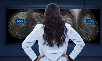
Revealing the breast's architecture
2D imaging - whether analogue or digital - is thought to miss detection of 20-30% of breast cancers. Early clinical results from studies using the new technology tomosynthesis indicate its potential to lower those percentages.
During a tomosynthesis examination, an X-ray tube moves in an arc around the breast, producing image slices that are virtually free of overlapping parenchyma, so manufacturers say this system can provide more accurate 3D views of the breast than the 2D views currently produced by mammograms. Tomosynthesis also delivers a lower radiation dose.
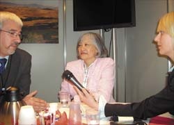
During the recent German Radiology Congress in Berlin, Roberta Agnes Jong MD FRCPC, head of the Breast Imaging Unit in the Medical Imaging Department of Sunnybrook Health Science Centre, Toronto, Canada, and José Abellan-Martinez, Marketing Manager for mammography in the Global Diagnostic Imaging division of GE Healthcare, discussed this firm’s tomosynthesis technology with Meike Lerner of European Hospital
‘Compared with today’s mammography, which provides us with a summation of images, where subtle abnormalities often get obscured by the images of other tissue, tomosynthesis will show us thin slices of the tissue of the whole breast,’ explained Dr Jong. ‘This will make it easier for the radiologist to look at the margins of masses, at architectural distortions and other signs of malignancy. So we hope that tomosynthesis will improve the early detection rate of breast cancer and will provide us with more accurate images that will reduce recall rates and the number of biopsies. This is a very important advantage for women, because a recall is always connected with fears and means great physiological and psychological stress. Furthermore, with the new method the breast must only be compressed one time and maybe with slightly less compression, but this is not proven at the moment. As a study has shown, the radiation dose during a tomosynthesis-examination is less or equal to that of a two-view mammogram.
‘For the radiologist, tomosynthesis will be a great help, for the analysis of the images will be much easier, because of their accuracy, and the danger of missing a detail will be minimised.’
Asked what advantages GE´s tomosynthesis equipment might have, compared with other similar technology in the works at other companies, José Abellan-Martinez said: ‘GE´s advantage is that we do have huge experience in the reconstruction of organs via CT, MR or RAD and we profit from our own technologies now regarding tomosynthesis. The technology we designed had all the features necessary for tomosynthesis, whereas other companies have to change their current technology used for digital mammography. So GE is one step ahead. A second point is, that our system is very efficient in terms of the radiation dose and image quality. But actually, we do not bother that much about other companies doing similar things; we have the right system and tomosynthesis is just another step to GE´s aim of early health. What is important in the end is that our work will result in a good, proven instrument that will support us in the fight against cancer.
‘Currently we are doing extended clinical trials that hopefully will prove our hopes. The first results will be available in the near future. Afterwards we need FDA approval. So, from my point of view, the first tomosynthesis systems will not be implemented before 2009 or 2010. But, as far as we know from other new innovations, it will be a long way before tomosynthesis will be the common method for breast cancer examinations. Just look at digital mammography: GE Healthcare started with its digital system in 1999. In 2006, only around 20% of all US hospitals were equipped with a digital mammography system. So today we cannot predict when women will benefit from this new technology. Hopefully it will be as soon as possible.’
20.06.2007