Germany's early detection programme and research
In 2002, Germany implemented an early detection programme for breast cancer. The digital Reference Centre For Mammography at the University Hospital Münster is one of five such centres in the country - and it's one of the most modern, providing digital systems for imaging and results evaluation as well as a mammo-PACS.
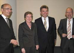
During a recent visit to Münster, Professor Andreas Pinkwart, Minister for Innovation, Science, Research and Technology of North Rhine-Westphalia, was updated on the screening programme by Professor Walter Heindel MD, who heads the Centre. They also discussed future diagnostic possibilities involving innovative imaging procedures. Present at the briefing, we asked Prof. Heindel about the programme’s success and particularly the research project focused on tomosynthesis.
In North Rhine-Westphalia the screening programme has been comprehensively implemented according to European guidelines, Prof. Heindel told us: ‘New digital systems have been installed in many surgeries and hospitals, which shows how seriously radiologists take this subject. The feedback is also positive among women entitled to be screened. In the first year more than 50% of women that the programme targeted (aged between 50-69 years) took up the offer of being screened. This great response initially surprised us, and now we can present first scientific data showing that we have been able to detect small, and therefore curable, breast cancer with the help of early screening. Our objective is to lower mortality from breast cancer through early diagnosis.
‘Being responsible for the mammography screening programme for North Rhine-Westphalia – Germany’s biggest federal state – the Centre is responsible for technical and medical quality assurance, training of screening teams. Our scientific focus is the evaluation of digital mammography technologies and of the programme. We work in multidisciplinary teams comprising radiologists, medical physicists, medical documentation assistants and radiology technicians, so that we assist the doctors participating in the programme at all times along with providing regular training to keep them up-to-date with the latest technological developments.
Currently you are also looking at new mammography technology - photon-counting tomosynthesis. Is this an inferior form of CT diagnosis?
‘Individual manufacturers are experimenting with a breast-CT. In terms of tomosynthesis, what we don’t need and want is for women to be examined by yet another machine. In fact, the idea is to use tomosynthesis only to further investigate suspicious results. We are currently co-operating with the Radiology Department at the Charité in Berlin in an EU-funded project looking at the kind of medical questions where tomosynthesis will be helpful, with the objective of comparing those results systematically with MR mammography. So far, MR mammography has been a problem solver for many women, such as those affected by lobular carcinoma, which is hard to detect with X-ray mammography. Its spread is hard to determine, because it often does not differ from the density of the normal glandular tissue. MR mammography is the gold standard here, prior to resorting to surgery. The question is whether we will be able to better detect this lobular carcinoma with the help of tomosynthesis. This is where the photon-counting system supplied by Sectra*, which significantly lowers the levels of scattered radiation, comes into the equation (see box). We have already confirmed this with our own measurements. However, the most important question - as always in imaging – is to what levels we can lower doses whilst maintaining the quality of diagnosis. That’s the decisive issue.’
Particularly because radiologists must resist the impulse to increase the dose to achieve brilliant images where a lot can be seen very precisely?
‘Yes, this is a well-known problem - a problem with digital technology such as this type of technology “evens out”, unlike conventional X-ray technology. Here you get over-exposed images when the dose is too high. This is the danger with digital mammography or generally with digital technologies. Theoretically the manufacturer or user can say: I want images of particularly brilliance and quality, i.e. I will increase the dose a little. But even if you still stay below the legal exposure limits when doing so, the interest of the women patients must be at the centre of things. The objective is to make that important diagnosis whilst adhering to the principle of protection from radiation, which is accepted the world over: as low as reasonably achievable (ALARA). This is a fundamental issue, which our working group is also looking into.
How do you know whether you just can’t see anything or whether you cannot see anything because the dose was too low?
‘This is a problem, which is why we currently work with so-called reference values, i.e. you state what dose should be used for an average examination. However, in digital mammography we have maximum limits that have been specified across Europe. I am convinced that, with the help of these new technologies, we can lower the doses and that’s why I have lobbied for this technology for our reference centre. Between brilliant images achieved with doses that are too high, and a situation where the doses are lowered to the extent that there is no diagnostic value left in the images taken, is where we need to define the right maximum and minimum values.
‘This includes the comparison of different digital technologies by collating relevant data. This is also the background for a request from the State Ministry to our reference centre to collate data on the doses administered, and to correlate them with the diagnoses, which will help to make realisable and scientifically well-founded conclusions. This is also of high interest from a political point of view, for instance in dealing with self-help groups for women.’
* Sectra manufactures diagnostic imaging equipment, including digital mammography systems. The firm reports that MicroDose Mammography is the only system that guarantees very low radiation doses for patients, and that the development of photon-counting tomosynthesis, with even lower radiation doses, is therefore a logical further development for the firm. Sectra adds that the system is currently only used for research purposes. The company is currently undertaking research in co-operation with, among others, Prof. Heindel within the EU research project ‘High Resolution X-Ray Imaging for Improved Detection and Diagnosis of Breast Cancer’.
The University of Münster
In Germany, Münster’s Medical Faculty is one of the top faculties of its kind for teaching and research. The secret of this success lies in the organ-centred and interdisciplinary orientation of the study courses. During any one semester students are lectured on a certain topic in all relevant medical fields, for example radiology, pathology, laboratory medicine or surgery, so they learn their subjects relating to certain indications. The results achieved by the students in their first state examinations - held according to the new, stricter guidelines - appear to justify our methods, the Faculty points out. ‘Münster students have done very well by comparison.’
A further strength in Münster is that innovative imaging, together with medical physics, constitute a defined platform at the Medical Faculty, and this will be further expanded. In this way, issues going beyond pure medicine, such as exposure to radiation, radiation measurements and safety issues can be swiftly evaluated.
20.06.2007