Mammography: What really counts?
By Prof Mats Danielsson, of the Royal Institute of Technology, Stockholm
The fact that the female breast is one of the most radiation sensitive organs in the human body is a major driver for all those searching for low radiation alternatives - one of these routes lies in photon counting.

At this year’s meeting of the Radiological Society of North America (See box), ‘Photon Counting: Is it the future of X-ray Imaging from Mammography to CT?’ will be an important panel discussion initiated by Professor Mats Danielsson of the Royal Institute of Technology, Stockholm, and Robert Nishikawa, Associate Professor of Radiology at the University of Chicago Medical Centre and Director of the Carl J Vyborny Translational Laboratory for Breast Imaging Research.
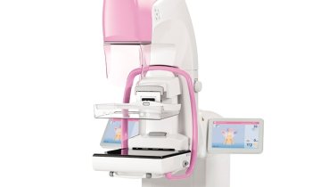
Is dose the only factor that determines image quality? The Swedish firm Sectra certainly does not think so. It claims that the Sectra MicroDose, currently in use in more than 10 countries, produces more brilliant images at half the radiation dose used by competing systems. We asked, Is this possible?
Prof Mats Danielsson explained: ‘The limitation of current imaging systems is that they have too much noise – comparable to a hearing difficulty in a noisy environment. Basically you have two options: either reduce the background noise or speak louder. To speak louder is equivalent to increasing a radiation dose; the only problem is that it puts women at increased risk for radiation-induced cancers. The other solution is to reduce the background noise, in a silent environment you can clearly hear a whisper, while even shouting may not help if surrounding noise is too high. In exactly the same way it is possible to achieve as good as, or, in fact, better image quality at half the radiation dose if just the noise is taken care of. With Sectra’s solution the main noise sources in terms of electronic noise and scattered radiation are reduced more or less to zero and this is really the answer. It’s not black magic.
‘This is achieved by photon counting technology. With the advent of applications such as tomosynthesis and dual energy mammography, it will become even more important to control noise because the signal in tomosynthesis will be much smaller compared to the noise – if nothing is done to reduce it. To count the X-ray photons down to the quantum level is the ultimate solution. Today, it is a significant advantage; it will be even more important in the future. Getting rid of the noise is the way to go.’
Apart from eliminating noise, it is also important to optimise the X-ray source in terms of target and filtering and this can be applied to all systems – photon counting or not. For example, some manufacturers still use X-ray tube anodes and filters similar to what was used for film (Molybdenum), which is obviously not wanted. However, since there is really nothing new in this area in the last decades, the potential for improvement is limited. The obvious choice is a detector without any noise combined with its optimum X-ray tube and filter.
A reduction in radiation dose may even be worth a slight increase in cost; in this case it is better to count the X-ray photons than just dollars and cents.
Photon counting
Since X-rays are digital and Sectra’s detector counts them one by one, a direct capture of individual X-rays occurs. This means no electronic noise in the image and no information loss in conversion steps, which is the case in other digital detectors. With Photon counting, there are no phantom images to interfere with interpretation, since the detector is fast enough to be ready when the next photon arrives. The image is acquired by a multi-slice scanning technology that eliminates the scattered radiation and significantly reduces the noise level in the image. The multi-slice scanning technology ensures that images are totally reliable, with no dead pixels that could obscure microcalcifications.
Photon Counting:
Is this the future of X-Ray imaging? Focus session at the RSNA, Monday 24 November 4:30-6:00 pm.
Professor Mats Danielsson says that 10 years from now all X-ray detectors for medical imaging will be photon counting.
Conversely, Professor Rüdiger Schulz-Wendtland, who heads the local mammography centre in Erlangen, Germany, questions: ‘Is photon counting really required? The radiation dose and image quality with traditional digital technologies are good enough for mammography’.
Photon counting detectors need to demonstrate benefits over other detectors prior to widespread acceptance, adds Lorens Niklason of Hologic Inc.
Their arguments and reasoning, along with those of Hans Ringertz (Stanford), Matthew Wallis (Cambridge), Robert Nishikawa (Chicago), Andrew Maidment (Philadelphia) and John Rowlands (Toronto), promise to make this debate lively indeed.
28.10.2008


