29th Annual Congress of the German Society for Senology
Competence for the benefit of women
Every year the German Society for Senology congress facilitates interdisciplinary breast cancer discussion between gynaecologists, radiologists, surgeons, pathologists, internists, radio-oncologists and plastic surgeons. In an interview with Karoline Laarmann, of European Hospital, radiologist Professor Ingrid Schreer (right), head of the Breast Centre at the University Women's Hospital in Kiel, and Deputy Chair of the Society, highlighted aspects of its 2009 congress this June

Welcoming the appointment of internist oncologist Professor Ulrich R Kleeberg as this year’s congress president, Prof. Schreer explained that he will place particular emphasis on individualised therapy, meaning a holistic, patient-focused treatment concept. ‘The responsible use of innovative treatment procedures will therefore be an important topic at the congress. What may technologically or pharmaceutically be possible may not always be the best choice for the patient’s well-being. However, the best possible quality of life, particularly for very young patients at high risk, is of enormous importance.’
The Certified Breast Centres project, run by the German Society for Senology and the German Cancer Society has met with initial success, and will again come under discussion during the congress. ‘They represent big progress for quality assurance,’ Prof. Schreer pointed out. ‘Through their guideline-orientated therapies, and the pooling of interdisciplinary specialists under one roof, the facilities provide the highest level of care for patients. However, the networking of the breast centres with the national mammography screening programme is not yet ideal. The DGS is hoping to encourage essential structural changes. It doesn’t make sense to offer screening — which has a high success rate in the detection of in-situ and small carcinoma – when, if a positive diagnosis has been established, the patient cannot be referred to a clinic with a certified breast centre that ensures appropriate medical care. A further shortcoming that needs to be overcome is the lack of comprehensive information and education for women. Unfortunately, information available about the risks and benefits of an early detection programme is currently only superficial. I feel it is particularly the gynaecologists who actively ought to seek discussion with their patients.
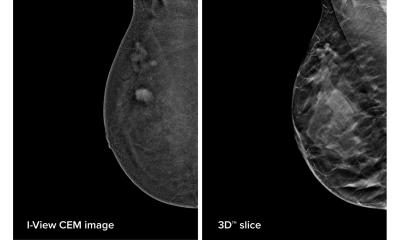
Asked about the promising potential of full-field mammography using tomosynthesis, which is currently being tested, the professor said she believes early screening with the established imaging procedures is ‘covered rather well’ and that ‘whatever new developments are added to these will have to find their niches.’
‘Theoretically,’ she added, ‘tomosynthesis appears promising because 3-D imaging can show the smallest areas of glandular tissue without overlay, but it remains to be seen what use this information about structure and composition of the breast will be in the end. Moreover, we don’t yet know whether this technology is as effective for the detection of microcalcifications as for tumour detection. As the use of two-dimensional mammography together with tomosynthesis increases the radiation exposure, I think it is best initially to rely on the standard procedures, i.e. ultrasound, for the examination of dense glandular tissue.’
3-D ultrasound
‘One focal point of the congress will be the topics around adjuvant hormone- and chemotherapy. The objective here is to shrink tumours larger than 2cm in size, so that it’s easier to operate on them and to preserve the healthy surrounding tissue. If the size of a tumour could be determined accurately with the help of 3-D ultrasound this would be of high prognostic importance. However, we need studies to evaluate how reliable the macroscopic ultrasound measuring procedure really is, by comparing the results with those gained through histopathological analysis. Unfortunately, there has not yet been enough research in this area.
Current trends in ultrasound
‘One interesting approach currently being studied in the USA is the rediscovery of scintigraphy. A higher resolution has been achieved through an improved generation of systems, which could become of use to detect breast cancer as well. As scintigraphy not only delivers anatomical information but also functional information it would be conceivable that it could be superior to MRI for breast examinations on dense glandular tissue. However, the basic problem with scintigraphy, i.e. being dependent on radioactive substances, which results in considerable radiation exposure, so far remains, even with the latest generation of detectors.
‘Moreover, there is currently a lot of experimentation with high fields in breast MRI to improve image quality. However, the first results show that although 3-Tesla can increase sensitivity, this happens at the expense of specificity. The danger of MRI then is that we get results that point towards a need for further clarification and investigation, which later on proves to have been unnecessary. Ultimately this is not in the interest of the patients.
01.05.2009


