Robot moves steadily in on catheter ablation
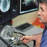
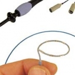
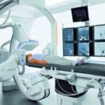
The Sensei Robotic Catheter System, a first generation robotic platform launched by Hansen Medical at the USA's Heart Rhythm Society Scientific Sessions in May this year, is in use in Europe.


The Sensei Robotic Catheter System, a first generation robotic platform launched by Hansen Medical at the USA's Heart Rhythm Society Scientific Sessions in May this year, is in use in Europe.
During a meeting of cardiologists in Prague earlier this year to exchange experiences with new methods and treatments to control atrial fibrillation, Dr Josef Kautzner, Head of Cardiology Department at IKEM (Institute of Clinical and Experimental Medicine) pointed out that numbers of patients with AF will more than double during the next 20 years.
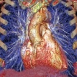
It has only recently been discovered that very often it is not the size of the plaque in the coronary vessels but its inflammation status that determines the occurrence of a cardiac infarction.
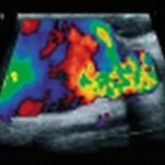
By Thaddeus Chodakauskas BS RDMS and Steve Feinstein MD FACC
Boston Scientific Corporation has commenced enrolment of a targeted 1,500 patients for the Taxus Perseus clinical trials, planned to take place in 100 international centres.
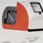
The Lifebridge B2T (bridge-to-transport) is the first, fully portable emergency life support system for patients suffering cardiogenic shock, or those showing signs of imminent cardiogenic shock.
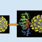
A next-generation diagnostic tool for cardiovascular disease, using a nanoscale iron particle, is now under development at a unique industry-government-university named Nano AG. A report from Siemens describes the research and progress at the centre
By Paolo Montuschi MD, of the Department of Pharmacology, Faculty of Medicine, Catholic University of the Sacred Heart, Rome, Italy.

The European Respiratory Society (ERS) Congress is the world's biggest annual scientific gathering in respiratory medicine.
Turkey - The recently opened 74-bed Florence Nightingale Kiziltoprak Hospital, which has three operating rooms and 10 ICU beds, is one of four hospitals in a Turkish healthcare network.

Trends 2007 - ever smaller and smaller
The Forum Medizintechnik-Pharma - an association that provides a recognised contribution to the development of the co-operative environment in medicine, technology and pharmaceuticals, met for a symposium in Germany this July.

"Come and experience the great Canadian Prairie Hospitality!" say the organisers of the 5th World Conference on Breast Cancer (WCBC), to be held in June next year in Winnipeg, Manitoba - the "cultural cradle of the nation, gateway to the Canadian west, and a meeting place for over 6,000 years," the WCBC Foundation points out.
A decision by Ab Klink, Minister of Public Health, Wellbeing and Sports, to increase the number of balloon angioplasty facilities in hospitals to 30, has prompted the NVVC - the Dutch cardiologists association - to express concern that there will be too many centres and too few patients, and specialists will not be able to maintain the level of skills for this procedure.
Cardiac CT angiography (CTA) performed after coronary artery bypass grafting surgery can reveal a high prevalence of unsuspected cardiac and significant non-cardiac findings that might otherwise be overlooked, according to a study by researchers at the University of Maryland Medical Centre, Baltimore ('Cardiac CT Angiography after Coronary Bypass Surgery: Prevalence of Incidental Findings', Pub:…
Hitachi Medical Systems has opened its European Technology Academy in Dusseldorf
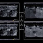
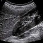
Ultrasound scanning with CCDS is an established technique in shunt diagnostics and allows non-invasive assessment of vascular flow. Stenosing changes to walls of vessels used as a dialysis shunt should be detected as early as possible to avoid occlusion by a thrombus. High occlusion rates with volume flow reduced by up to 45% in one year demand ultrasound screening. The risk of haemodynamically…
Despite efforts in recent years to reduce the number of deaths caused by breast cancer it is still the most common cancer occurring in women. Approx. 47,500 new cases of the disease appear in Germany per year, a situation comparable with other European countries and with the USA. In 2004 alone, 17,592 women died from the sequela of a breast carcinoma (Federal Statistics Office statistics on…
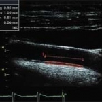
Ever heard this from a patient? 'Wouldn't it be great if it was in colour.' It's true; B mode lacks the glamour of Doppler. We're guilty of it ourselves, we love Doppler. How frequently do we hear 'did you put colour on that'? When was the last time you asked 'how much temporal smoothing did you use'? And yet every time I watch others scan I witness Sonographers scanning from various angles,…
In recent years MR angiography techniques have steadily created new possibilities in patient studies. The lack of radiation exposure and the fact that these new techniques are non-invasive, or at most minimally invasive, are just two of the obvious benefits. And in particular, within the last few years contrast-enhanced MR angiography has become an ever more preferred tool. This requires…

Ischaemic stroke accounts for 80-85% of all cerebrovascular accidents and causes considerable morbidity and mortality, thereby placing a significant burden on western societies. In the UK alone, stroke costs amount to 10.4 billion EUR annually. These events commonly are a consequence of systemic atherosclerotic disease and with the internal carotid arteries supplying 75% of cerebral blood flow,…
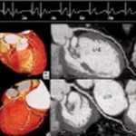
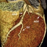
The advent of multislice computed tomography has made coronary artery imaging with computed tomography (CT) a clinical reality. When a CT scanner with 16 detector rows is used, scan times are within the breathholding capabilities of most patients. The latest generation of CT scanners with 64 detector rows has reduced the scan time to 6-10 seconds, which is suitable for examining all but the most…
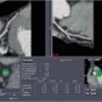
Ongoing refinement of modern helical multi-slice CT (MSCT) scanners offers the opportunity for very high temporal and spatial resolution thin-slice data set acquisition. The reconstruction techniques offered (multisegment reconstruction) result in improved temporal resolution down to 40-200 ms and thus also reducing motion artifacts. The SURECardio software package makes possible optimum studies…
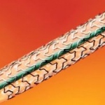
Coronary stenting is the most common way of treating symptomatic ischaemic cardiopathy and stenosis of venous bypasses. Restenosis actually occurs in 30% of patients treated with non-medicated stents while "drug eluting" stents have reduced the incidence of restenosis by approximately a quarter and have proved particularly effective in diabetic patients and in the treatment of lesions…
Radiotherapy treatment planning relies on transversal CT images. They form a basis for treatment planning, dose calculation and increasingly the plan localization for external radiation therapy, called CT simulation. A dedicated CT scanner for radiotherapy plan simulation is one of the most essential pieces of equipment in a modern radiotherapy department. In contrast to external beam…