CT Perfusion of the Brain

The basics of the method and interpreting images

CT assisted dynamic perfusion imaging (perfusion CT, PCT) has evolved in recent years with the introduction of the multi-slice spiral technique, the use of study protocols with lower injection rates and improved evaluation programmes.
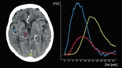
Fig. 1: Typical time/density curves after injection of a contrast mediumbolus...
Fig. 1: Typical time/density curves after injection of a contrast mediumbolus in Perfusion CT. The density sequences are imaged (idealised view) in an arterial vessel (middle branch of the cerebral artery: blue), a venous vessel (confluence of sinuses: yellow) and in the cerebral parenchyma (thalamus: red). Note the typical staggered time between the arterial and venous time/density curves and the flattened
and slightly delayed density sequence in the parenchyma compared with the arterial curve

This article was first published in the VISIONS, issue 9/2006, a publication of Toshiba Medical Systems

03.07.2007

More on the subject:More on companies:

Related articles

Photo

Sponsored • Computed Tomography

Mobile Imaging Solutions: Clinical Confidence Anywhere

To bridge new equipment installations in radiology, temporary high workloads, or long-term additional capacity, Canon Medical provides mobile solutions according to customers’ needs and wishes.

Photo

Video • Redesign & refresh

Canon Medical updates Aplio ultrasound systems

Canon Medical announced the commercial launch of the Aplio i-series / Prism Edition, a complete redesign of its premium ultrasound series. In addition, Aplio a-series, Canon Medical’s…

Photo

Sponsored • All-in-one imaging trailer

Mobile solutions for the COVID-19 frontline

With public health issues continuing to make daily mainstream news headlines across the world, it is clear how much change the healthcare environment is going through. Not only are there existing…

Related products

Subscribe to Newsletter