Sonography in Renal Disease

Ultrasound examination of the renal tract is one of the primary imaging methods to verify the presence of urinary obstruction, calculi, neoplasms, and other focal findings. This short article summarises important aspects of sonography for some typical renal abnormalities without claiming to be a comprehensive treatment. Since sonography is frequently nonspecific, clinical laboratory findings are of utmost importance in the assessment. Consequently, the ultrasound examination should be undertaken by the clinician.

Fig. 1: Normal-sized right kidney of a 65-year oldmale with smooth surface,...
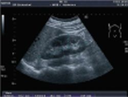
Fig. 1: Normal-sized right kidney of a 65-year oldmale with smooth surface, homogenous echo pattern of echogenicity less than the liver and typical echo-poor medullar pyramid

This article was first published in the VISIONS, issue 9/2006, a publication of Toshiba Medical Systems

12.07.2007

Read all latest stories

Related articles

Photo

Sponsored • Computed Tomography

Mobile Imaging Solutions: Clinical Confidence Anywhere

To bridge new equipment installations in radiology, temporary high workloads, or long-term additional capacity, Canon Medical provides mobile solutions according to customers’ needs and wishes.

Photo

Video • Redesign & refresh

Canon Medical updates Aplio ultrasound systems

Canon Medical announced the commercial launch of the Aplio i-series / Prism Edition, a complete redesign of its premium ultrasound series. In addition, Aplio a-series, Canon Medical’s…

Photo

Sponsored • All-in-one imaging trailer

Mobile solutions for the COVID-19 frontline

With public health issues continuing to make daily mainstream news headlines across the world, it is clear how much change the healthcare environment is going through. Not only are there existing…

Related products

Subscribe to Newsletter