MRI
Metamaterials boost sensitivity
A group of researchers from Russia, Australia and the Netherlands have developed a technology that can reduce Magnetic Resonance Imaging (MRI) scanning times by more than 50 percent, meaning hospitals can drastically increase the number of scans without changing equipment. This extraordinary leap in efficiency is achieved by placing a layer of metamaterials onto the bed of the scanner, which improves the signal-to-noise ratio.

The details of this experimental research are available in the current issue of Advanced Materials. This patent-pending technology is currently being co-developed by MediWise, a UK based company that specializes in commercializing metamaterials for medical applications.
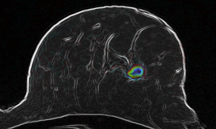
MRI is one of the key methods of modern diagnostics that have found wide applications in medicine, biology and neurology. MRI scanning helps monitor the subtlest of physiological changes in our internal organs. For instance, a timely MRI procedure can detect tissues affected by cancer in the earliest stage of the disease. The possibility of effective MRI diagnostics, however, depends almost entirely on the quality of resulting MRI images.
Scientists from ITMO University, Australian National University, Ioffe Physical-Technical Institute, University Medical Center Utrecht and Institute of Experimental Medicine RAMS demonstrated that the quality of MRI images could be substantially increased with the aid of metamaterials - artificial periodic structures that can interact with electromagnetic radiation in an extraordinary fashion.
"This is the first real demonstration of the practical potential of metamaterials for MRI imaging enhancement and scanning time reduction. Our research may evolve into new healthcare applications and commercial products." - says Yuri Kivshar coauthor of the study, head of the Nonlinear Physics Centre at the Australian National University.
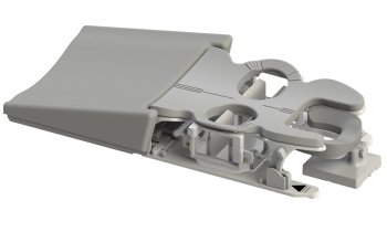
By placing a specially designed metamaterial under the studied object in an MRI scanner it is possible to increase the signal-to-noise ratio in the scanned area. The result of this increase is that either a higher resolution image can be obtained over the same time slot or faster examination can be performed with the same resolution as in an ordinary MRI scanner. In addition, the metamaterial suppresses the electric field, which is responsible for tissue heating - a phenomenon that may compromise the safety of the whole MRI procedure.
The problem of tissue heating has recently become even more relevant with the arrival of high-field and ultra-high-field MRI scanners in the medical practice. A drive for high-field MRI is mediated by the benefits of better image resolution However, tissue heating becomes substantial at higher fields due to an increase of the radiofrequency energy absorption. Therefore, the issue of safety in high-field and ultra-high-field MRI scanners remains open.
The scientific group managed to entirely avoid tissue heating, at the same time preserving high resolution. The solution, in essence, does not require any intervention into the hardware of the MRI scanner, but rather represents an inexpensive functional add-on device that can be used with any existing MRI scanner.
"Our metamaterial can be embedded directly into the patient table of any commercially available MRI scanner. However, in the future we see even more potential in the concept of special smart clothing for MRI scanning," says Alexey Slobozhanyuk, first author of the study and researcher at the International Laboratory of Applied Radiophysics at ITMO University. - "Stripes of our metamaterial can be sewn in the clothes. The examination of patients, wearing such clothes, would lead to higher resolution MRI images, while the special design will enable a homogeneous enhancement of the signal-to-noise ratio, which does not pose any risk to the patients' health. As a result, with metamaterials you will be able to improve the characteristics of low-field MRI to the extent that their functionality is comparable to high-field MRI."
The duration of an MRI exam also presumes inconveniences for patients. In ordinary MRI devices, the scanning may last from 15 to 60 minutes and during this time the patient must remain completely immovable. The possibility of achieving detailed images in a shorter time slot will make the procedure more comfortable for the patient and in the long view could even reduce queue time in hospitals.
"Our idea of using metamaterials in order to receive images with higher detalization will allow doctors to localize and study oncological diseases. Based on the images obtained with an MRI scanner, the surgeon determines the structure of the inflammation, which afterwards will serve as a blueprint for his scalpel during the operation." - says Yuri Kivshar.
"Metamaterials have been proven to add value through their ability to process electromagnetic and sound waves in ways that no natural material can do. This leads to emerging business opportunities creating genuinely disruptive products. The scientific field of metamaterials in rapidly evolving and impacting traditional industries such as Aerospace, Telecoms, Cleantech and now Healthcare , " - concludes George Palikaras, Mediwise's Founder and CEO. "The technology has the potential to extend the life of MRI imaging machines but more importantly, it will make the scan quicker, more accurate and safer to patients. We are honoured to work alongside world leading academic partners, and to help advance this important innovation from the laboratory to the marketplace"
Source: ITMO University
15.01.2016


