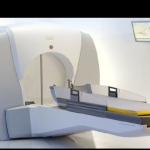
The Gamma Knife goes to work in Paris
A major public hospital has become the first in Paris to be equipped with a Gamma Knife, the device that enables the surgeon to operate on the brain with no blade or blood involved.



A major public hospital has become the first in Paris to be equipped with a Gamma Knife, the device that enables the surgeon to operate on the brain with no blade or blood involved.
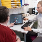
PredictAD is an EU-funded research project that develops objective and efficient methods for enabling earlier diagnosis of Alzheimer's disease. Diagnosis requires a holistic view of the patient combining information from several sources, such as, clinical tests, imaging and blood samples.
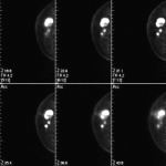
Every medical congress is an opportunity for the manufacturers to showcase their products. This year’s congress of the German Röntgen Society was no exception -- and one innovation particularly caught the attention of our European Hospital team: positron emission mammography, PEM for short.
CT-PET is the child of a forced marriage between nuclear medicine and radiology. A shared session at ECR 2011 in Vienna did little to assure there is a growing consensus between the two partners.

Although developments in molecular imaging still do not reach the high expectations placed upon them, the change in imaging is very obvious. Having been limited to the imaging of morphology, nowadays information on tissue characteristics, blood vessels or the metabolic behaviour of tumours provide many more insights, for example into response behaviour.
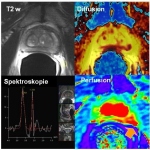
Thinking of the future of imaging, inevitably PET-MRI springs to mind. The fascination of this novel hybrid technology is great, seeing how it combines the best from three imaging areas: anatomy, function and metabolism. The further development of functional procedures in oncology is raising particularly high expectations. However, how extensive the use of this potentiated image information will…

Molecular Imaging (MI) emerged in the early 21st Century as a discipline at the junction of molecular biology and in vivo imaging to enable the visualisation of the cellular function and the follow-up of the molecular processes in living organisms. Modalities available for MI encompass MRI, CT and ultrasound, PET, as well as Optical Imaging, and are by nature frequently experimental.

A leading radiologist is forecasting a ‘paradigm shift’ in breast imaging. Dr Peter Brader, from Department of Radiology, Division for Molecular and Gender Imaging, Medical University Vienna, envisages that diagnosis and treatment will move from a ‘one size fits all’ approach to one of personalised molecular medicine by 2025. He also foresees greater use of theranostics with combinations…

Whole-body hybrid PET-MR scanners are emerging on the market and are expected to have a significant impact in diagnostic imaging particularly in oncology applications and also in other clinical domains, such as cardiology, inflammatory and infectious disease, as well as in neurological applications.

"One size fits all" – the phrase is a fact of life in terms of the drugs available to treat cancer patients today. This solution can bear tragic results. Only 25% of cancer patients currently respond to this ‘one size’ drugs administration. In addition, 100,000 patients die annually, in the USA alone, from the side effects of those drugs. Personalised therapies that are devised to suit…
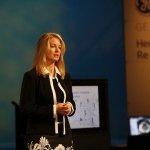
When planning radiotherapy the combination of positron emission tomography (PET) and Computed tomography (CT) can provide a better outcome than CT alone. Michael Krassnitzer asked Terri Bresenham MSc BSc, Vice President for Molecular Imaging at GE Healthcare, for her views on the value of PET/CT, the new EANM guidelines, novel tracers and the future of other hybrid imaging technologies.

High resolution radionuclide imaging is a technique increasingly used to detect breast cancers and has already been shown to offer improved diagnosis in many clinical situations. The technique, which will be discussed at RSNA 2010 (28 November to 3 December, Chicago) , is also allowing clinicians to detect previously unknown areas of breast cancer in women with newly-diagnosed disease.

Late in 2009, GE Healthcare’s 64 slice PET/CT Discovery 690 began its use in the University Clinic for Nuclear Medicine in Innsbruck, Austria. Apart from routine clinical examinations for treatment control via FDG (fluorodeoxyglucose), the scanner is also employed in clinical research - particularly for Gallium 68DOTA PET/CT examination.
Molecular imaging, the discipline that unites molecular biology and in vivo imaging technologies to assess biological activity in the body, promises to open up ‘…an entire new universe,’ declared Dr Ralph Weissleder, of the Centre for Molecular Imaging Research at Massachusetts General Hospital, USA, in the journal Radiology. That was just one decade ago. And he was right. It has indeed…
2010 workshop in Aachen, Germany, in March, were welcomed by Professor Thomas M Deserno, head of the Medical Informatics Department at RWTH Aachen University and organiser of what turned out to be an ‘outstanding’ scientific programme.

In the current economic environment the introduction of novel imaging approaches and their reimbursement by payers is becoming increasingly difficult. Historically, this denotes a significant change, in that many currently accepted routine tests or interventions were accepted based on common sense, convincing experience or rapid adoption into clinical routine without much scrutiny.
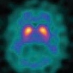
Over the past decades new imaging technologies have substantially broadened the range of imaging applications in clinical medicine. For years anatomical imaging modalities, such as X-ray and CT, reveal high-resolution information of organs and tissues over extended imaging ranges. Lately, however, the idea of functional imaging e.g. the visualisation of physiology in vivo gains importance.
Molecular imaging, the discipline that unites molecular biology and in vivo imaging technologies to assess biological activity in the body, promises to open up ‘…an entire new universe,’ declared Dr Ralph Weissleder, of the Centre for Molecular Imaging Research at Massachusetts General Hospital, USA, in the journal Radiology. That was just one decade ago. And he was right. It has indeed…

Clinicians gathering in Brussels for the first-ever conference on Molecular Imaging in Radiation Oncology (MIRO), from 18-20 March, will take the first steps toward setting practice guidelines for using PET scans to target radiotherapy, a task that will culminate with publication of these recommendations in September.

Molecular imaging, a new science that emerged from molecular biology, is unlike traditional imaging. Whilst the latter can, for example, show the differences in proton density or water content on MRI, molecular imaging uses biomarkers (probes) that interact selectively with molecules within an area and then generate the image according to fine molecular alterations occurring inside (e.g. within a…

A novel molecular imaging technology aimed at rapid diagnosis of cell death in organs such as the brain and heart has been licensed by The Medical College of Wisconsin, USA, to GE Healthcare. The method uses imaging probes with a radiopharmaceutical compound. Under the license GE will further evaluate and develop the technology and will have an option to commercialize it.
In the last fifteen years, myocardial perfusion SPECT imaging (MPI) has been performed most commonly by dual-head conventional scintillation cameras with parallel-hole collimators, configured in a 90ºdetector geometry and image reconstruction based on standard filtered back projection (FBP) algorithms. Such arrangement, although clinically well established suffers from important limitations…
Founded and managed by Prof Peter Schlag, the Charité Comprehensive Cancer Centre (CCCC) co-ordinates cancer diagnostics and therapies across all medical disciplines. Interdisciplinary tumour boards decide on therapies/strategies; cooperation is close with general practitioners, regional hospitals and clinics.
Bayer Schering Pharma AG, Germany, has presented positive data on a global Phase II study with the novel positron emission tomography (PET) tracer florbetaben (BAY 94-9172) at the International Conference on Alzheimer's Disease (ICAD) in Vienna, Austria. This study showed that patients with a clinical diagnosis of Alzheimer´s disease could be differentiated from age-matched healthy volunteers…
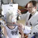
Although the potential of proton therapy was recognised over half a century ago, and since its development is now known to deliver a radiation beam accurately into a tumour without damaging surrounding tissue, high equipment costs limit its general introduction. Mark Nicholls reports on a British hospital with two decades of experience in its use - and value

