What's new in PET technology?
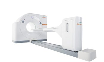
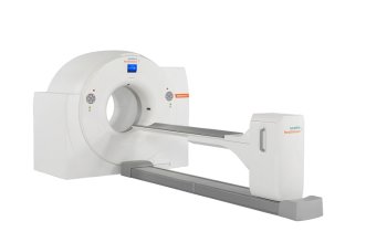
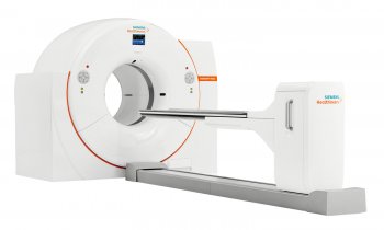
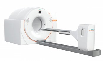
Positron emission tomography (PET) is a fancy tool for metabolic imaging. This week Tom Lewellen from the University of Washington Medical Center in Seattle takes a detailed look on what is to expect of the recent developments in PET detector technology.
Positron emission tomography (PET) is a tool for metabolic imaging that has been utilized since the earliest days of nuclear medicine. A key component of such imaging systems is the detector modules—an area of research and development with a long, rich history. Development of detectors for PET has often seen the migration of technologies, originally developed for high energy physics experiments, into prototype PET detectors. Of themany areas explored, some detector designs go on to be incorporated into prototype scanner systems and a few of these may go on to be seen in commercial scanners. There has been a steady, often very diverse development of prototype detectors, and the pace has accelerated with the increased use of PET in clinical studies (currently driven by PET/CT scanners) and the rapid proliferation of pre-clinical PET scanners for academic and commercial research applications. Most of these efforts are focused on scintillator-based detectors, although various alternatives continue to be considered. For example, wire chambers have been investigated many times over the years and more recently various solid-state devices have appeared in PET detector designs for very high spatial resolution applications. But even with scintillators, there have been a wide variety of designs and solutions investigated as developers search for solutions that offer very high spatial resolution, fast timing, high sensitivity and are yet cost effective. In this review, we will explore some of the recent developments in the quest for better PET detector technology.
To read the full study from Physics in Medicine and Biology please click on the following link:
26.08.2006


