1st 64-slice combined PET-VCT scanners go to work
Greater diagnostic accuracy predicted for cardiology, oncology and neurology
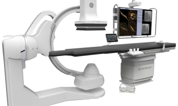
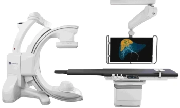
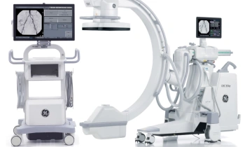
During the annual meeting of the Society of Nuclear Medicine, in California this June, GE Healthcare announced the first installations of its new Discovery VCT - described as “the world's first true 64-slice combination positron emission tomography and volume computed tomography (PET/CT) system.”

This combines the Discovery Dimension platform, the high-speed and high-resolution provided by GE’s volumetric CT, with the high sensitivity motion imaging capabilities of its Discovery PET system - a combination of technologies that should enable physicians to more accurately diagnose and identify heart disease and cancer and neurological disorders etc.
Along with an installation in the USA, GE installed Discovery VCT in the molecular imaging research institution Turku PET Centre, in Finland, where Professor Juhani Knuuti, the centre’s director, said: ‘PET and VCT imaging allows linking the anatomical findings of coronary arteries with the information of myocardial perfusion, function and metabolism. That may improve the accuracy of the assessment of myocardial viability. However, the greatest potential of Discovery VCT lies in future applications of molecular imaging, matched with precise anatomical detail in imaging the coronary arteries we will be pursuing, using the tools provided in Discovery Dimension motion PET imaging capabilities.’
Daniela Zimmermann, of European Hospital, spoke with Professor Knuuti about the scanner and its predicted potential for cancer and cardiac diagnoses.
‘Our previous PET-CT (Discovery STE) produced some very nice oncology applications - of course, the major indication of a PET-CT. Now, with a fast multi-slice PET-CT we can expand usage to cardiac applications, particularly for coronary diseases,’ Professor Knuuti explained.
As I understand it, PET scans nuclides moving through the body - a procedure that takes time, whether combined with a 16-slice or 64-slice CT. Does the number of CT slices really matter?
‘Yes. With a 64-slice CT we can perform a high quality coronary angiography in about five heartbeats - that’s roughly six seconds. With the fast multi-slice PET-CT complete cardiac perfusion studies are fused with a high quality CTA in one single examination that lasts under 30 minutes. Moreover, the system presents us with the possibility of working on future applications, such as imaging vulnerable plaques and stem cells.
A 16-slice CT needs more than half an hour?
‘The difference is that, with a 64-slice CT, you get the entire patient cardiac angiography in five heartbeats when the heart is in a stable phase, resulting in an unsurpassed image quality. With a 16-slice CT (or 32-slice CT) it takes longer and might require more patient preparation, and the success rate is lower.’
Then the 64-slice CT volume is fused with the PET data?
‘Yes. For perfusion studies you typically start with the CTA. For normal cases - i.e. no stenosis in the coronary angiogram - thanks to the high negative values, you can conclude that the patient has no coronary disease and stop here. But, very often when you study patients with more likelihood of cardiac disease you will get positive findings, such as calcified plaques, which are difficult to interpret. These findings required, before the Discovery VCT, another cardiac investigation. Now we can immediately perform cardiac PET studies and link the artery disorder with the lack of perfusion. In one single examination we can provide a complete assessment of cardiac functionality.’
Commonly PET-CT is used in oncology, to locate tumours. Has the new scanner has changed that usage?
‘The staging of cancer is still the main use, because you can find metastasis that will influence treatment decisions. Since the development of advanced cardiac applications, on both the PET and CT sides, interest in using the PET-CT in cardiology becomes more obvious.’
Does this mean diagnoses are more accurate?
‘Clearly, the Discovery VCT give us more confidence in our diagnoses. Cardiac application in PET-CT will become increasingly important with the development of new PET agents that will help in characterising coronary plaques, for example. In the Turku PET Centre, we are testing a couple of new tracers to image inflamed plaques, which are likely to rupture (the so-called vulnerable plaques).Without this new, hybrid system we could not reach that target.’
01.07.2006