Role of New Software and Technology
Albert Flotats Giralt
In the last fifteen years, myocardial perfusion SPECT imaging (MPI) has been performed most commonly by dual-head conventional scintillation cameras with parallel-hole collimators, configured in a 90ºdetector geometry and image reconstruction based on standard filtered back projection (FBP) algorithms. Such arrangement, although clinically well established suffers from important limitations including long image acquisition, low image resolution, and patient radiation dose.
The quality of the reconstructed SPECT images depends on that of the acquired images, which is affected by (a) photon scatter and absorption (physical effects that are particularly important in MPI because the variety of densities surrounding the heart), (b) acquisition geometry (variablespatial resolution), and (c) patient motion.
Novel reconstruction algorithms include modelling of physical phenomena and acquisition geometry, increasing contrast and decreasing noise of acquired images, which enables shortening of acquisition time on conventional gamma cameras by a factor of ≥ 2 preserving image quality. In addition, optimisation of imaging systems with dedicated detectors and collimators for MPI improves photon detection efficiency and spatial resolution. The combination of both new reconstruction algorithms and new imaging systems results in much higher photon sensitivity and improved image quality and resolution, allowing acquisition times as short as 2 minutes. These new developments facilitate new imaging protocols with improved patient comfort, increased throughput, and reduced patient radiation dose.
New reconstruction algorithms
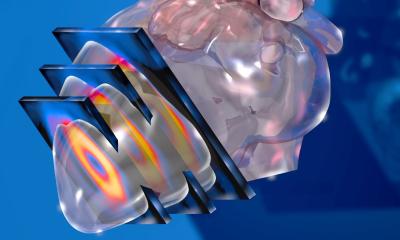
SPECT reconstruction is based on the generation of the 3-dimensional distribution of a radiotracer from a series of 2-dimensionalimages (projections) acquired at a sufficient number of positions around the object being imaged, generally along an arch spanning at least 180°.
Back projection is an analytic solution to tomographic reconstruction. The activity in a given pixel in a projection is projected backward (backprojected) onto the pixels in the distribution that are in its direct path. Linear superimposition of backprojections from different angle views produces the tomographic image but generates a “star” artefact. FBP partially solves this artefact by filtering each backprojection with a high-pass filter (ramp filter). This process is associated with image noise amplification, which is reduced by applying a low-pass (or smoothing) filter toprojection images before reconstruction, at expenses of loss of resolution and contrast. FBP reconstruction assumes that distribution of a radiotracer is detected equally in all of the angular projections, leading to various artefacts caused by variations in attenuation, scatter, resolution, and count density.
Currently, improvements in computer power have made possible other reconstruction approaches based on algebraic calculations that also use projections to find the distribution of activity in the field of view. Since the exact solution to this problem is not possible when matrix dimensions are not very small, an approximated iterative method is used. Iterative reconstruction initially guesses the value of allpixels using FBP, which is subsequently slightly altered several times(iterations) until meeting an appropriate final result (2). Iterative reconstruction facilitates the incorporation of compensation algorithms for physical photon effects into the reconstruction process reducing artefacts (photon scattering from extracardiac activity, photon absorption from soft-tissue attenuation and variation of spatial resolution as function of depth).
Different proprietary iterative algorithms based on maximum likelihood expectation-maximisation (MLEM) orordered subsets expectation maximisation (OSEM) have been developed. MLEM use sprojection data from all radial angles in each iterative estimation step, whereas OSEM groups projection data into an ordered sequence of N subsets(typically 2-4 projections per subsets) for 4-12 iterations, using only 1/N of the total data at each iteration, decreasing computing time without measurable loss of image quality. Reconstruction image data are updated for each subset during each iteration. Consequently, the number of updates is the product of iterations and projections subsets. As the number of updates increases, the spatial resolution increases, at expenses of increasing image noise. Therefore ,most current algorithms use various forms of noise suppression during iterativere construction. A proper balance between the number of iterations, the number of subsets and noise smoothing filter is needed in order to obtain optimal image quality (3).
A different reconstruction development that has recently shown to improve MPI quality is the ‘‘motion-frozen’’ processing of gated SPECT images, which eliminates blurring due to cardiac motion with analgorithm that shifts counts from the whole cardiac cycle into the enddiastolic position. The resulting images have the appearance of end-diastolic frames but are significantly less noisy since the counts from the entire cardiac cycle are used. The spatial resolution of such images is higher than that of summed gated images (4).
New imaging systems
Conventional gamma cameras derive from Anger’s invention in 1957 and are based on transparent sodium iodide crystal detectorcontaining thallium impurities -NaI(Tl)- coupled to photomultiplier tubes(PMT). Recently, new dedicated MPI systems with improved detector materials and/or geometry and improved collimator design have been introduced by various vendors, improving tomographic sampling of the myocardial region.
Silicon photodiode or solid-state detectors offer the potential for improved spatial and energy resolutions, with greater stability than conventional detectors. Combination of improvement in spatial resolution and sensitivity allows faster imaging times.
Some of these new systems perform SPECT imaging with the patient in a (semi-)/upright position, improving patient comfort and reducing claustrophobic sensation, further reducing patient motion. Another advantage refers to space saving since this equipment is more compact than the conventional one. Different systems with digital logic signal processing are commercially available. Digirad® provides 2- or 3-solid-state head detectors consisting of pixilated CsI(Tl) crystals coupled to individual silicon photodiodes, with prospect to market a system which includes an ultra low-dose x-ray transmission source for subsequent attenuation correction. CardiArc is based on a rotating thin curved lead-sheet collimator with 6 narrow verticals lots and additional stationary lead vanes stacked vertically facing3 curved stationary NaI(Tl) crystals coupled to 3 rows of PMT (20 PMT perrow). D-SPECT® consists of 9 vertical columns of 1024 CZT pixilated CZT elements each, fitted with square, parallel hole, high sensitivity tungsten collimators, placed in a 90º gantry geometry.
Other vendors have explored image collimation using multi-pinhole design, which allows many views to be acquired simultaneously throughout the entire image acquisition period without the need for motion of the detector, collimator or patient, which eliminates view-to-view inconsistencies and reduces artefacts induced by patient motion. In addition, multi-pinhole collimation design does not need electro-mechanical hardware, potentially reducing the manufacturing and servicing costs. However, potential limitations of multi-pinhole design include (a) generation of artefacts due to the inherent production of incomplete tomographic dataset and image acquisition from only limited views (5); (b) inconsistencies in the reconstructed data dueto extracardiac activity may not be seen by all of the views; and (c) decrease of the resolution and sensitivity of pinhole collimators with the distance from the object being imaged (although this can be compensated when resolution recovery is applied in the reconstruction).
Further development of solid-state and semiconductor detectors will strengthen their performance and cost-effectiveness, and eventually replace conventional gamma cameras.
References
1. Hesse B, Tägil T, Cuocolo A, Anagnostopoulos C, Bardiés M, Bax J, et al. EANM/ESC procedural guidelines form yocardial perfusion imaging in nuclear cardiology. Eur J Nucl Med Mol Imaging2005;32:855–97.
2. Germano G. Technical aspects of myocardial SPECT imaging. J Nucl Med 2001; 42:1499-1507.
3. Slomka PJ, Patton JA, Berman DS, Germano G. Advances in technical aspects of myocardial perfusion SPECT imaging.J Nucl Cardiol 2009;16:255-76.
4. Slomka PJ, Nishina H, Berman DS, etal. ‘‘Motion-frozen’’ display and quantification of myocardial perfusion. JNucl Med 2004;45:1128-34.
5. Funk T, Kirch DL, Koss JE, BotvinickE, Hasegawa BH. A novel approach to multipinhole SPECT for myocardial perfusionimaging. J Nucl Med 2006;47:595-602.
27.10.2009