POCI: an intra-operative imaging probe for sentinel node localisation
By Stéphanie Pitre PhD, co-inventor of the TreCam camera, CNRS, Paris
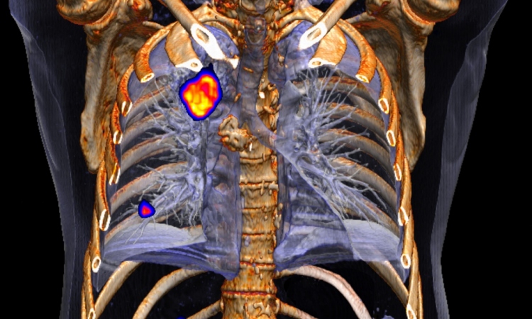
Growing interest in sentinel lymph node (SLN) detection has led to its use in many clinical centres. The most successful protocols for localisation of SLNs are those that include preliminary lymphoscintigraphy performed with a conventional gamma-camera. In the absence of a nuclear medicine department, SLN protocols using only an intra-operative counting probe are generally less efficient. To overcome this problem our group has developed a high resolution intra-operative imager, POCI (Per-Operative Compact imager) dedicated to nuclear medicine use.

of the Imagerie et Modélisation en Neurobiologie et Cancérologie laboratory, UMR 8165, University Paris 7-Denis Diderot and University Paris-Sud 11
The current hand-held POCI prototype is based on a 40 mm diameter intensified position sensitive diode (IPSD) coupled to an interchangeable gamma head module. The spatial resolution of the POCI camera is 2.2 mm FWHM (in contact), a corresponding detection efficiency of 10.7 cps/µCi and an energy resolution of 32%. The total weight of the POCI device is 1.2 kg and the corresponding outer dimensions are 95x95x85 mm3.
Since January 2006, a clinical study (N°P040417) at the Tenon Hospital (Paris) has been evaluating its use in a sentinel node localisation protocol for breast cancer staging in 200 patients. A peritumoural injection of 160 MBq 99mTc labelled nano-colloids is performed two hours before the lymphoscintigraphy, which is realised pre-operatively, first using a conventional single-head gamma-camera and then the POCI camera. These examinations are carried out by two different operators blinded to each other’s results. The POCI is used in contact with the patients’ skin scanning the whole axillary area with 10-second acquisition images.
The following day, the surgeon uses POCI to perform an intra-operative lymphoscintigraphy and then, the counting probe for transcutaneous SLNs pre-localisation. The number and the localisation of SLNs obtained by both intra-operative detectors are compared with those obtained the day before with the conventional gamma-camera. So far, 105 patients have been included in this clinical protocol. All SLNs initially localised by the standard gamma-camera were detected with the POCI camera within exploration times that ranged between two and 10 minutes.
These preliminary clinical results show that the POCI camera is able to predict the number and localisation of all SLNs.
16.11.2007