Automatic Quantification of Left-ventricular Function
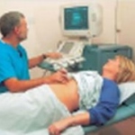
The evaluation of the size and function of the left ventricle in patients with suspected heart disease is a central diagnostic problem. In contrast to other methods of evaluating left-ventricular function (e.g., angiocardiography, right-sided heart catheterization, radionuclide ventriculography or MRI), transthoracic echocardiography is a widespread and readily available procedure that is not…