New technology can reduce number of biopsies
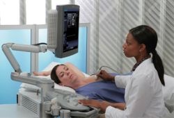
Approximately 75 percent of all biopsies are negative, which is of course the good news. But looking at the side effects of this invasive method combined with a long waiting time for the results, a biopsy is nevertheless an unpleasant procedure for the women affected. A new promising technology now offers the possibility to differentiate benign and malignant tissue due to an adjunct of the regular breast ultrasound examinations and therefore could reduce the number of biopsies.

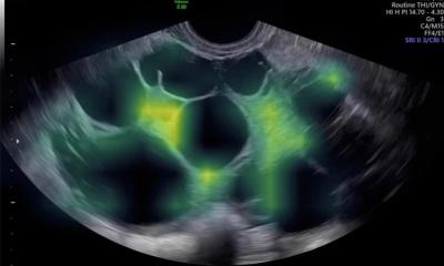
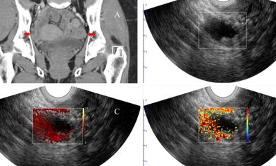
“eSie Touch Elasticity Imaging”, so the name of the new technology is a software recently introduced by Siemens Medical Solutions and available with the 5.0 release of the Acuson Antares ultrasound system. With the new application, physicians can generate an “elastogram” which provides additional information about mechanical properties, e.g. the stiffness of breast lesions. The method offers a significant improvement in the acquisition of data – in general, the heart beat and the breathing of the patient will provide a sufficient movement to generate an elastogram.
Currently the method is being tested in several studies with promising success. In a recent published American study, 80 patients with a total of 123 suspicious lesions were examined using the elasticity measurement. 18 lesions were classified as malignant, which was confirmed in 17 cases by a biopsy. Of the 105 lesions predicted as benign, all were biopsy-proven benign.
The results are presently being validated in comprehensive studies in Europe.
Background
Elasticity imaging illustrates the relative stiffness of tissue compared to its surroundings. As tissue undergoes pathologic changes, its relative stiffness will change. The stiffness of the tissue as well as its size compared to the B-mode image provides further insight into potential pathology.
Of course, the ability to visualize tissue elasticity could not replace biopsies in general, but there is a reason for hope that this method may reduce the number of unnecessary breast biopsies.
29.03.2007
More on the subject:


