3D USCT nears reality
Back in the 70s, when scientists first speculated on the development of 3-D ultrasound computed tomography (3-D USCT) the available technology could not equal their dreams. Now, before the end of 2007, a prototype at Germany's Karlsruhe Research Centre will be used for the first in-vivo tests.
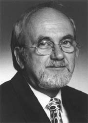
EH reporter Meike Lerner asked Professor Hartmut Gemmeke, Head of the Institute for Data Processing and Electronics, at the Centre, and one of the developers of 3-D USCT, why such a prototype has taken so long to create, and what it might achieve
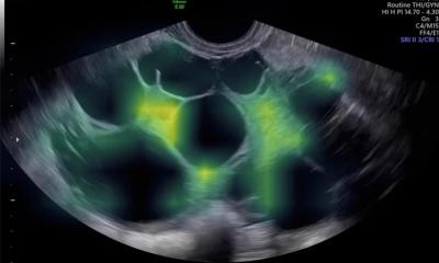
“Our prototype currently works with 1,600 ultrasound sensors – implementation with that many sensors, using conventional sensor technology, would have been too expensive. So, first we had to develop effective but cheap sensor technology. Then again, the amount of data generated by a 3D ultrasound CT image is a problem. In mammography, the amount of data we receive, per image per breast, if left uncompressed would fill 32 CDs. Reconstructing this image from the measured data would take an average PC about a month. That’s where we were dependent on the development of appropriate algorithms and hardware. Around five years ago we finally realised that technology had advanced to a stage where it was feasible to contemplate the idea of 3D USCT again.
Now we are at the stage where we have carried out the first tests with nylon threads of 0.15mm, which were very successful. By the end of the year we will be able to test the sensitivity of this method for mammography in the first in-vivo tests.
The conditions are promising: On the one hand we’ll be able to screen the breast from all sides, so therefore avoid shadowing effects. On the other hand the breast is not squashed, so that we will be able to locate growths and tissue changes in the 3D image as well. We expect to be able to detect tumours of less than 5mm. Moreover, this method will deliver reproducible images, which will also facilitate functional diagnostics. Compared with conventional ultrasound, 3D USCT not only measures reflections and reductions but also the speed of sound and frequency shifts. Because of the many sensors that are directed towards one picture element, the speckle noise typically produced with ultrasound scanning is extremely reduced, which is a further advantage.
Therefore, 3D ultrasound CT offers possibilities to show all modalities together in one image – and this means we’ll probably be able to obtain information about the disease pattern of breast cancer at a molecular level. We’ve carried out first examinations with tracers, which can then be seen in 3D via ultrasound; however, this method is still in its infancy and we cannot yet say much about the sensitivity. However, if our expectations are confirmed, this would mean we’ll be able to detect and quantify breast cancer in its very early stages.
Therapeutic use via hyperthermia is a further vision we have for 3D USCT. The conditions for this are given due to the large number of ultrasound sensors that focus as actors and can shell the tumour using hyperthermia.
However, these are dreams of the future. Initially we’d like to prove, with clinical tests, that 3D USCT does indeed have the effects in mammography for which we hope.
Therefore we plan to find a partner in the medical industry who will help us to implement this technology - we expect this will take two to three years. Then it will probably take another two years before the first equipment reaches hospitals. By the way, the price for this type of equipment is likely to be similar to what we currently pay for digital mammography equipment.
20.06.2007