Article • Organ donation
The liquid biopsy
Donor transplant rejection and cancer relapse have two things in common: early recognition is vital and monitoring is hugely challenged.



Donor transplant rejection and cancer relapse have two things in common: early recognition is vital and monitoring is hugely challenged.
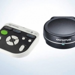
Olympus has released a new standalone controller for the 5 megapixel DP26 camera. Using the controller, it is possible to view samples and capture images directly on a monitor screen with no need for a dedicated computer.
Research in the field is booming thanks to newly arriving methods to identify gene sequences. Scientists are interested in a wide range of issues from disease-relevant variations of human genetic information to the detection of viral genetic material that supports therapies. Several highlights of current research were presented this spring at the 9th International Symposium on Molecular…

This May, IFCC WorldLab Berlin with its manifold scientific programme gives clinical lab physicians the opportunity to see over the rim of the tea cup of their working field. Within the congress theme ‘Healing the world’ comes an exciting lecture on laboratory work in third world countries and disaster areas.

A new technique to aid the early detection and diagnosis of inflammatory bowel disease has been developed by UK and Germans researchers. The Confocal Laser Endomicroscope (CLE) contains a powerful microscope that allows clinicians to view bacteria in the intestine that are thought to trigger bowel diseases such as Crohn’s and ulcerative colitis.
Clues to the causes of serious, and often fatal, diseases and conditions, such as heart disease, stroke and high blood pressure, are being investigated at the Universities of Strathclyde and Glasgow, both in Scotland, with the use of a £1 million grant.
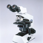
SECARS microscopy, a procedure which is based on quantum effects, with which certain molecules can be monitored virtually in real time, turns out to be a highly sensitive tool for the non-invasive research of the most varied diseases and may supplement the medical diagnosis tools of CT and MRI in the future.
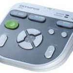
The new Olympus DP21 digital microscopy camera for high-definition (HD) image capture is suitable for use in life science and materials microscopy. The 2.11 megapixel stand-alone camera allows easy observation, focusing and framing, while enabling the smooth display of real-time 1600x1200 (UXGA) images, the manufacturer reports.
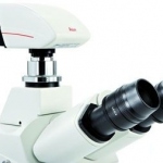
The detection and documentation of low light fluorescence signals in live cell experiments is a particular challenge for digital cameras. To meet this demand, Leica Microsystems adds the new Leica DFC345 FX to its portfolio of powerful digital cameras. In addition to combining high sensitivity with high resolution, this new camera features a fast image capture rate and a broad dynamic range,…
"In-vitro veritas", the slogan of the 18th European Congress of Clinical Chemistry and Laboratory Medicine, which took place in June, underlined the significance of in vitro diagnostics for healthcare, and the presentations by 90 speakers, in 32 scientific sessions and 29 industry sponsored workshops, demonstrated the rich variety of developments.
Medical products have to fulfil exacting requirements. That is why surface properties are increasingly analysed with optical 3D measurement systems.

Since the completion of the 13-year human genome project in 2003, molecular scientists have been delving deeper into the world of cells.
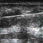
Early suspicions of cancer, either from clinical evidence or medical imaging, are often followed up on biopsy. However, the removal of tissue for histological analysis can be uncomfortable for patients.
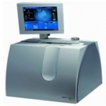
Recently in February, Leica Microsystems, Wetzlar, Germany, acquired Bal-Tec AG, Liechtenstein-based manufacturer of optical instruments. This transaction will allow Leica to expand its product portfolio for EM (electron microscopy) specimen preparation significantly.
Europe's biggest microscopy, imaging and analysis event
Opening up new dimensions in ergonomics and mobility — in close cooperation with leading neurosurgeons, Leica designed a new swivel arm for its M525 OH4 surgical microscope that offers 36% more reach than comparable instruments.
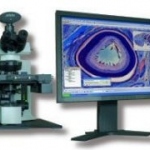
The updated Olympus dotSlide digital virtual microscopy system can scan entire slides at high resolution and fidelity, making them accessible and fully navigable anywhere in the world. Using any of the three models - dotSlide MD (manual); dotSlide SL (fully automated, with slide loader), and dotSlide TMA (with a tissue micro-array module) - users can examine a virtual slide as if seeing the…
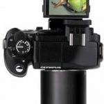
The new Olympus E-330 micro-imaging system for microscopy includes the world's first digital SLR camera to show real-time frame images on the LCD, the company reports. `The system is based around a 7.5 Megapixel sensor, which together with an array of unique features delivers incredibly sharp and vibrant images directly onto a 2.5 inch high-resolution colour LCD. Other features of the micro…
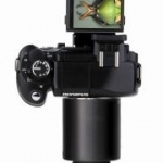
The Olympus E-330 digital micro-imaging system promises new perspectives for biological procedures: With the unique digital Single-Lens Reflex (SLR) camera real-time frame images can be shown on a LCD (Liquid Crystal Display).
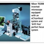
Bio-imaging Facility at the University of Manchester ordered three new Nikon eC1 confocal microscopes.