Live cell imaging
Bio-imaging Facility at the University of Manchester ordered three new Nikon eC1 confocal microscopes.
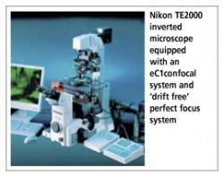
Two of these are to be equipped with EMCCD monochrome digital cameras, offering single photon sensitivity. Nikon reports that one of the confocal systems will be attached to a Nikon Eclipse 90i upright microscope, whilst the others will be fitted to TE2000E inverted motorised microscopes, making them ideal for a wide range of advanced live-cell imaging techniques. These inverted microscopes will also be equipped with Nikon’s perfect focus system (PFS) to eliminate drift during time-lapse observations. ‘The PFS has to represent one of the most useful additions to any live cell imaging microscope,’ explained Dr Peter March, Bio-imaging Experimental Officer at the Faculty. ‘Contrast-based auto focus is just not an option in rapid live cell imaging. For TIRF, maintaining a stable focal plane is critical - any drift during the experiment will make the data meaningless. With PFS, once you set that focal plane it stays in focus and doesn’t move.’
17.11.2006