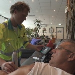
Video • Emergency care
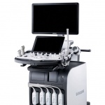
Point-of-care ultrasound helps save time and lives
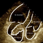
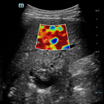
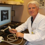
Time is of the essence in an emergency situation, and may be the difference between life and death. Ambulance crews on the front line must decide rapidly whether or not a patient is suffering from a life-threatening condition requiring specialist treatment, and point-of-care ultrasound can provide vital guidance. Geert-Jan Deddens, a nurse practitioner in emergency care with the Rotterdam…