Article • Remote robotics
Long distance ultrasound is not so distant
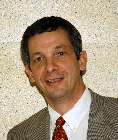
The future of medicine lies at a distance, underlined Professor Michel Claudon, Head of Radiology at the Regional University Hospital, Nancy, France, whose particular interest is ultrasound.
Advances in IT and telecommunications have driven expert healthcare provision even into the most remote rural areas – underlined by this notable test in ultrasound.
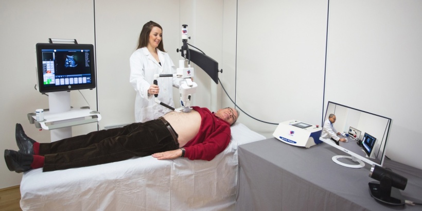
The Melody System is the world’s first remote ultrasound imaging system. Produced by French company AdEchoTech, and developed by radiologists for radiologists, the system has evolved to meet the real needs of an ultrasound examination. Recognising that the necessary precision would be impossible using only a voice-controlled robot, the system incorporates the most recent audio-visual teleconference technology with advanced remote robotics similar to those used in space exploration.
The lead radiologist controls the entire examination from a central workstation, where the video link enables the patient, operator and ultrasound images to be seen on high-resolution computer screens throughout the examination. The highly sensitive audio-link also enables the expert radiologist to instruct the operator and discuss with the patient throughout the procedure.
However, most importantly, it is the use of a ‘dummy’ probe by the expert that drives the robot and therefore, ensures exam quality. While watching the images, the expert moves the dummy probe as they would if physically at the patient’s side.
Currently used primarily for abdominal ultrasound examinations, the lead radiologist has complete control over the set-up parameters of the images, such as gain control, depth control, frequency change and activation of various ultrasound modes, such as Colour Doppler and Pulse Wave Doppler.

Is the system as good as it seems? Claudon’s team organised a test to find out. The Melody system was set-up in the Lunéville Hospital centre, over 30 km away from the specialist centre in Nancy.
The equipment was very easy to set-up – within a day it was completely operational in the Lunéville ultrasound room with the secure audio-video link picking-up the images in Nancy.
Over the next three days approximately 30 different abdominal ultrasound examinations were performed with the system. Following instructions from Nancy, the patients placed themselves on the couch and operator positioned the robot over their body and applied gel. The probe on the robot, inclined at 45° then followed the movements of the expert’s dummy probe to perform the examination.
The team were quite surprised at how quickly they adapted to using the system, within two-three examinations ‘to use a dummy probe honestly became completely natural’. Also the images provided are completely compatible with the hospital’s PACS, so they could be automatically archived for future reference.
What about the patient? In this series of tests, patients were completely relaxed and compliant and had absolutely no difficulty with the concept of having the specialist at a distance. For them the end result of the examination is the same but without the loss of time and other potential difficulties of having to travel to the specialist centre.
Are there limitations? The system is as good as the internet connection for the specialist to feel that they are present at the examination. It has to be performed in real time, with no pauses and screen break-up. While fibre-optic cable will produce the best results, for longer distances satellite connections may add up to a millisecond of difference, which nevertheless should be imperceptible to the radiologists concerned.
There is also the ergonomic question of probe depth because the pressure exerted by the robotically controlled probe is not the exact equivalent of the radiologist’s hand but, as with robotic surgery, future developments and greater use of such systems will overcome.
The team in Nancy are unanimous in praising the system and can see advantages far outweigh the minor disadvantages. They consider it could enable specialist radiologists to intervene earlier in diagnoses, reduce travel for frail patients, training of junior radiologists and a host of other applications.
* European Hospital would like to acknowledge the help of Dr Alix Martin-Betaux, Dr Frédéric LeFevre (CHRU Nancy) and Dr Eric LeFebvre (AdEchoTec) in producing this article.
11.11.2016


