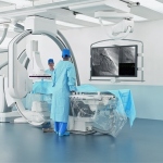
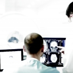
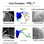
Siemens re-engineers radiography with flat emitter, new detector
In the highly competitive US market for interventional radiology, Siemens introduced a ground-breaking new imaging chain for the Artis line that sets a new standard for image quality.

In the highly competitive US market for interventional radiology, Siemens introduced a ground-breaking new imaging chain for the Artis line that sets a new standard for image quality.

The new Aquilion ONE ViSION is the widest, fastest, thinnest-slice CT ever built, capable to pushing both anatomical and functional studies to new levels.

To treat heart problems, common sense says we should look to the heart. New European research based on an advanced ultrasound system suggests that stiffening of the arteries plays a key role

Artificial hearts, originally designed to bridge the time on the waiting list for a heart transplant, in recent years have increasingly become an independent treatment option for patients with chronic heart failure (HF).

Ultrasound is enthusiastically embraced by cardiologists in guidelines as essential for evaluating a patient's heart. Now visualisation of 3-D wall motion takes ultrasound to a higher level, opening a new understanding of heart mechanics

A new generation of cardiovascular implantable electronic devices (CIEDs) includes the Implantable Cardioverter Defibrillator (ICD), Cardiac Resynchronisation Therapy Defibrillator (CRT-Ds) and Cardiac Resynchronisation Therapy Pacemakers (CRT-Ps). Professor W R Bauer at University Hospital Würzburg has been significantly involved in their development, EH Editor Ralf Mateblowski to ask him about…

More anatomy details, real-time visualisation of catheter movement, and reduced exposure – MRI has promising potential in rhythmology, explain Professor Matthias Gutberlet and PD Dr Christopher Piorkowski, at the Heart Centre, University Hospital Leipzig, Germany.

Over the last five years the tiniest particles have attracted large attention in relation to the diagnosis and treatment of cardiovascular disease. Indeed, as in other medical disciplines, nanotechnology is advancing in cardiology despite as yet insufficient research on the extent of its effect and double blind studies to confirm findings

John Brosky reports on a ground-breaking trial and how CT-FFR may change the practice of invasive cardiology and cardiac surgery.

Cardiovascular diseases, the most common cause of death in the West, includes diseases for which early detection is an important objective in cardiac imaging – particularly for coronary artery stenosis. Diagnosis is often made in the cardiac catheter laboratory. Now, however, CT scanning advances provide a lower impact alternative to that invasive exam. PD Dr Thomas Schlosser, Consultant at the…

Incorporating coronary CT angiography (CCTA) into the initial evaluation of low-risk patients coming to hospital emergency departments (EDs) with chest pain appears to reduce the time patients spend in the hospital without incurring additional costs or exposing patients to significant risks. The report of a study conducted at nine U.S. hospitals appears in the New England Journal of Medicine.

So close yet so far away! A fitting description of many diagnostic examinations of heart and lungs. Modern imaging modalities allow these two neighbouring organs to be evaluated together, nevertheless it is rarely done.

CVD prevention experts will meet in Dublin from 3-5 May for the leading international forum for preventive cardiology, Finn Snyder reports.

The Impax CV12A, a new version of Agfa HealthCare’s Cardiovascular Information System (CVIS), was introduced this March during the American College of Cardiology’s (ACC’s) 61st Annual Scientific Session & Expo in Chicago, USA.

EIZO GmbH, Display Technologies introduced the RadiForce LX600W, a large widescreen LCD color monitor with an LED (light-emitting diode) backlight. This 8 megapixel monitor is designed for use in operating rooms (interventional radiology, cardiology, and surgery).

They examine the structure of the heart muscle with magnetic resonance imaging (MRI) or evaluate the status of the coronary vessels with computed tomography (CT): radiologists increasingly use imaging methods to prevent or to assess cardiac diseases.

During the RSNA 2011, Professor Uwe J Schoepf MD, was asked what will be the chosen procedure of the future in cardiac imaging, he answered without hesitation: ‘Definitely CT,’ and, the Director of Cardiovascular Imaging at the Medical University Charleston, South Carolina.

Much has changed for medical device manufacturers. Take scanner development; whereas the aim has long been to increase multi-slices, produce higher field strengths and sharper images, optimise the ergonomics and then launch the new product at a specific group of customers, in recent years this approach became insufficient.

Medical imaging is no longer limited to diagnosis and evaluation. New, high resolution imaging systems enable very precise positioning of lasers, endoscopes and micro-instruments for surgery near the nerves, spinal cord or blood vessels and for selective tumour destruction, with minimum levels of pain and low complication rates.

With transcatheder aortic valve implants (TAVI) forming some 20% of all heart valve replacement procedures today, and the technology constantly developing, the 'real art' to the intervention's success lies in precise patient selection and procedure performance carried out by a multi-disciplinary and effective team, according to Simon Redwood, Professor of the interventional cardiology at King's…

The multipurpose BRANSIST alexa, which aims to provide total support for advanced catheterisation procedures, features a 12 x 12-inch flat panel detector (FPD) – an ideal size, the manufacturer Shimadzu points out, for covering interventions from head-to-toe, from brain blood vessels, cardiac and abdomen to peripheral blood vessels in the upper and lower extremities.

Prof. Uwe Nixdorff from the European Prevention Centre, Düsseldorf advocates cardiologists combine IMT measurement with ALOKA’s pulse wave intensity function to check for unseen coronary heart disease: “This technique is currently seldom used, however in my experience it provides a more complete picture and enables me to treat patients earlier for life-threatening conditions that are often…

Researchers in Sheffield have been given an mportant grant to trial state-of-the-art computer modelling systems that could provide a breakthrough in the treatment of patients with heart failure.