Aortic valve replacement in the third dimension
Meike Lerner reports
No white lab coats anywhere; instead men in hard hats, equipped with hammers and drills. The Düsseldorf University Hospital’s Cardiology Pneumology and Angiology Clinic is a construction site, but once the workmen have packed up their tools and removed the scaffolding the view to the human heart will be unobstructed and clearer than ever before. Here, innovative patient care and a highly ambitious research project are in the making. Buzz word: Hybrid.


‘In Düsseldorf, when we talk about hybrid, we talk about three things: First, there’s the hybrid operating room, which means that the spatial preconditions are met that specialists of different medical disciplines - cardiology, vascular and cardiac surgery - can treat the patient jointly at one OR table,’ explained Dr Jan Balzer, assistant medical director at the Department of Cardiology, Pneumology and Angiology ath the University Hospital in Düsseldorf. ‘Secondly, and this is the logical conclusion of the hybrid OR, there is hybrid intervention and, finally there is the really exciting area of hybrid imaging.’
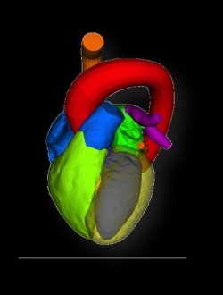
Hybrid imaging is nothing less than the fusion of image data from different modalities into a 3-D model of the heart. This will take place in a hybrid room (still part of the construction site) where MRI, rotational angiography (XperCT), fluoroscopy and 3-D echocardiography will be possible. ‘This room is the foundation of a research project that we will be conducted jointly with Philips and which entails the development of IT-based image fusion,’ Dr Balzer added. ‘The aim is to merge the advantages of each modality into one complete image of the heart. This would allow us to plan interventions, for example an aortic valve replacement, in a very detailed fashion and thus significantly reduce the risk. In a second step, haemodynamic data of the left ventricular outflow tract as well as ECG data will be added.’
The exact visualisation of the heart and heart valve structures before and during valve interventions is becoming increasingly important because the age of the cardiac patients is increasing. Most patients suffer from co-morbidities that make surgery more difficult, or even impossible. Planning transfemoral or transapical catheterisation for older, multimorbid patients with severe heart valve stenosis can be a highly complex task. But the different modalities contribute their specific advantages: CT visualises the anatomy of the outflow tract very well, transthoracic and transoesophageal live 3-D echocardiography offer an overview over the morphology of the heart valve. Angiography provides details on the coronary arteries and ventricle function can be evaluated with the help of cardiac MRI. Hybrid imaging will merge all this vital information into a 3-D model of the heart. Since it offers an image of the individual patient, it will allow fast and comprehensive identification of the patient’s individual risk profile prior to the intervention as well as planning the intervention itself.
The team around Prof Dr Malte Kelm knows the advantages of 3-D visualisation very well. In 2007, they were among the first in Europe to use the newly introduced and innovative Philips probe for 3-D transoesophageal echocardiography – not only for imaging but also for intervention. The 3-D technology is very well-suited for both diagnostics and therapy, for example in the visualisation of the aortic valve structure. Since the heart can not only be shown in 3-D but also in real-time, the physician gains a perfect overview of the region of interest.
Today, the transoesophageal live 3-D probe is a standard feature of minimally invasive interventions. Be it mitral valve interventions, percutaneous ASD or PFO occlusions – the procedures can be controlled far more precisely. ‘Several studies confirmed that the 3-D probe reduces both the rate of complications and examination time,’ said Dr Balzer, an experienced 3-D echocardiography user. ‘Moreover, 3-D echocardiography requires a much smaller radiation dose – a fact from which, above all, pregnant women and children benefit. The potential of 3-D transoesophageal echocardiography in cardiothoracic surgery has not yet been fully exploited.’
The 3-D technology available today and enormous progress in imaging technologies paved the way for the development of individual anatomical organ models. In the Düsseldorf research project the team is developing software that allows the simultaneous fusion of data from cardiac MRI, rotational angiography (XperCT), fluoroscopy and 3-D echocardiography to 4-D imaging in a patient-specific heart model. Complex image recognition procedures are used to locate pre-defined points of orientation and to identify the borders between different kinds of tissue. This novel hybrid imaging technology could become reality within the five-year research project. The long-term objective of the Düsseldorf team is the additional integration of molecular imaging data into hybrid imaging processes with the result of a tailored therapy for every individualized patient.
Image Courtesy of Dr. Jürgen Weese, Philips Research, Germany
30.08.2010


