


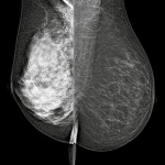
Study suggests a new method to measure breast density can help determine cancer risk

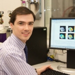
Dr Peter Choyke, Chief of the Molecular Imaging Programme at the National Cancer Institute in Bethesda, USA, believes that new tracers will have an evolving role to play and represent an exciting development in the imaging of cancers.
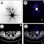
Although most nuclear medical examinations using SPECT (single photon emission computed tomography) take place beyond hospitals, two to three times more SPECT exams than PET-CT exams are carried out within hospitals.
Much, if not even everything, may have been said already about the multimodal approach in breast diagnostics. However, Professor Rüdiger Schulz-Wendtland at the Institute of Radiology, University Hospital Erlangen, says there is still surprising news from this field – innovations in multimodal breast diagnostics, for example.
UK researchers are working on a new MRI technique called hyperpolarised MRI – or Dynamic Nuclear Polarisation (DNP) – that can utilise more of the available nuclei than traditional MRI, helping to overcome some of its limitations by increasing sensitivity 10,000-fold or more. DNP is part of a longer-term aim to improve cancer mortality with the help of novel cancer imaging tools.
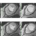
Today, magnetic resonance imaging receives top billing in cardiology next to the co-star computed tomography while much hailed single-photon emission computed tomography (SPECT) plays but a minor role.

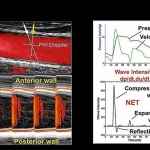
To treat heart problems, common sense says we should look to the heart. New European research based on an advanced ultrasound system suggests that stiffening of the arteries plays a key role

The first International Day of Radiology (IDoR) will be celebrated on November 8, as participating societies from all over the world will host a series of events to highlight the role played by radiology in modern medicine and help raise the public profile of the radiologist.
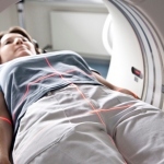
– The demand for multi-slice computed tomography (CT) systems will drive growth in the European CT market.
Global brands increase market-focus for photonics event and help investors to better spot potential investments in hardware.
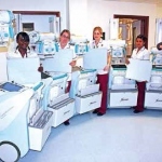
Conceptualized specifically for high-acuity patient monitoring and integrated with the world’s leading technologies, Edan Instruments’ latest elite V8 modular patient monitor offers real-time, organized view of accurate patient information, intelligent data analysis, management, network connectivity and simplified operation to the ICU, CCU, NICU, OR and PACU.

Demand for health services in China is on the surge, and democracy pre-empts an upward trajectory.

MIR is a subcommittee of the European Society of Radiology (ESR), focused on all aspects of management, which help to “Make Imaging Relevant” in today´s healthcare. Join our conference, which will take place in the exciting and busy city of Milan (Italy) and will last two full days from Thursday, October 11 to Friday, October 12.

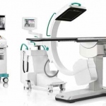
CT-ultrasound fusion optimises planning and monitoring of ablations in interventional radiology.
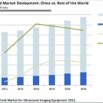
Market experience is likely to sharpen between local Chinese suppliers and multinational suppliers.

The use of contrast agents in ultrasound imaging of the gastrointestinal tract is no longer limited to the liver, although the clinical results of hepatic applications continue to be those that are most comprehensively confirmed.
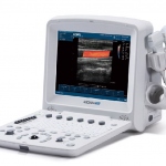
From China: The U50 portable colour imaging system