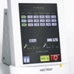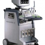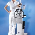
An accumulator-free contrast agent injector
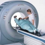
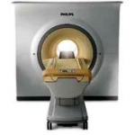
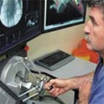
ulrich medical, established 25 years ago, is an independent medium-size medical technology firm with worldwide sales. Among its products are contrast agent injectors for contrast agents used in computer and magnetic resonance tomography.