
News • Research
Deaths higher for heart attack patients at night and weekends
Off-hour presentation and outcomes in patients with acute myocardial infarction: systematic review and meta-analysis


Off-hour presentation and outcomes in patients with acute myocardial infarction: systematic review and meta-analysis
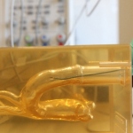
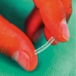
The Plaque-CharM project funded by the German Federal Ministry of Education and Research is to develop novel sensor technology that can characterise arterial tissue in the smallest space – the tip of a catheter.
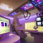
Prevention Suite’s four combined imaging technologies enables practical pre-clinical assessment of cardiovascular disease risk .
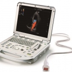
Okan Ekinci, Global Director of Cardiology at Siemens Healthcare, is convinced that, ultimately, ultrasound will remain the ‘entry level’ imaging procedure for patients. European Hospital met up with him at this year’s ESC congress to hear his thoughts on the potential of ultrasound – and particularly its fusion with other imaging modalities.

Performing manual chest compressions well for an extended period of time is almost impossible.

Football authorities across the world have been urged to adopt a universal standard of emergency care to help cut the potential for serious injury or death during matches.
Adding high quality, dynamic ultrasound for hybrid imaging enables clinicians to improve detection of a range of lesions or to intervene better for improved clinical outcomes. ‘We can no longer be fascinated with pictures; what we need is proof of the clinical benefit from tools and techniques,’ said Professor Jose Zamorano MD, Director of Cardiology at Ramón y Cajal University Hospital in…
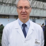
Part of the University Hospital Centre at Charleroi, the cardiology service provides consultations for a cluster of other hospitals, polyclinics and private physicians, which means that Dr Kathleen Retailleau takes to the road several days of each week to see patients throughout the region.
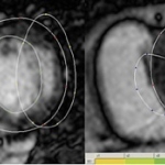
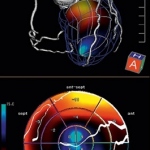
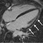
A cardiac catheter is insufficient to evaluate the effects of a myocardial infarction. The size of the infarction and post-event cardiac muscle activity are crucial predictive parameters that determine therapy decisions
Professor Axel Haverich and team at the Clinic for Cardiothoracic, Transplant and Vascular Surgery in Hanover Medical School (MHH) have been carrying out research into decellularised heart valves for over 15 years. They trialled a procedure – initially in the laboratory and in animal experiments – which does not cause tissue rejection, is hoped to last a lifetime and, in the case of children,…

We must not forget that despite advances, cardiovascular disease remains the number one killer in Europe.
‘Cardiology is one of the most innovative medical disciplines. Many modern technologies, such as catheterisations or imaging procedures, were triggered by cardiology,’ declared Professor Dr Gerald Maurer MD.
Ultrasound expands its role in cardiac imaging with disruptive applications. Fasten your seat belt. Cardiac diagnostics is entering a zone of turbulence. Manufacturers of leading systems continue to mine data from the sonic signal that opens new fields for research. John Brosky reports
Cardiologists believe they can restore coronary arteries thanks to a new generation of stents that help the body to strengthen collapsed vessels. Elsewhere, patients’ own stem cells are being programmed to rebuild cardiac muscle in HF patients.
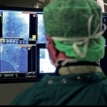
The Catharina Hospital (Eindhoven, the Netherlands) and Royal Philips (NYSE: PHG, AEX: PHIA) today announced the results of a clinical study involving the treatment of 136 patients with complex heart rhythm disorders such as atrial fibrillation (AF).

Computed tomography (CT) is the modality of choice for many diagnostic issues. Whilst currently its major strength is the visualisation of anatomical detail, future technological improvements may also reduce radiation exposure.
The potential of cardiac magnetic resonance imaging (CMRI) is still largely untapped. One novel application might be ablation follow-up. The first MRI-guided cardiac interventions were performed at Herzzentrum Leipzig, but, as far as coronary imaging is concerned, MDCT remains superior to MRI

Researchers in Germany have suggested that, for certain patients, newly developed coronary CT angiography techniques can provide good quality images with very low dose radiation.

The 2013 ESC Guidelines on Cardiac Pacing and Cardiac Resynchronization Therapy¹ developed in collaboration with the European Heart Rhythm Association (EHRA), have created a new classification system for bradyarrhythmias according to mechanisms rather than aetiology.

Smoking increases the risk of heart disease and stroke by five-fold in people under the age of 50 and doubles risk in the over-60s.

More than half of Germany’s population aged between 18 and 74 years cannot show off a gapless set of teeth, and that’s similar in France and worse only in Poland, according to a 2012 study, which also investigated oral hygiene.

Numerous cardiac muscle cells die following myocardial infarction, due to reduced blood flow in the affected muscle areas. What remains is a scar, which also mechanically affects cardiac pumping. The muscle itself has no, or hardly any, capacity to regenerate itself.
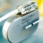
Not such a rare situation: A patient is due for an MRI scan to clarify a diagnosis. However, it transpires that this patient is fitted with an implant, say an implantable cardioverter defibrillator (ICD), which is contraindicated for MRI examinations.

How the different advanced cardio vascular imaging technologies fit together in managing cardiac patients, will be one of the main themes explored at the International Conference on Nuclear Cardiology and Cardiac CT (ICNC 11).
CT scanners now nicely cover morphology. The challenge is moving to CT functional imaging without frying patients