Nanotechnology
Over the last five years the tiniest particles have attracted large attention in relation to the diagnosis and treatment of cardiovascular disease. Indeed, as in other medical disciplines, nanotechnology is advancing in cardiology despite as yet insufficient research on the extent of its effect and double blind studies to confirm findings
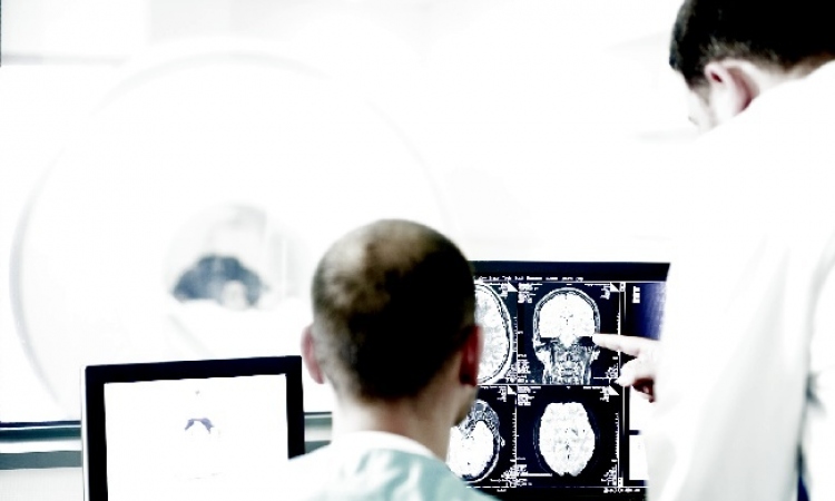
In cardiology, nanotechnology has two foci: Nanoparticles improve the early detection of arteriosclerosis in diagnostic imaging and are also used to deliver stem cells and medication to specific body areas with pinpoint accuracy. With standardised imaging, an arteriosclerotic vessel can only be detected once a morphological -- i.e. structural change, generally a narrowing (stenosis) of the lumen -- is already present. With conventional contrast media an infection of the arterial walls, which occurs in the early stage of arteriosclerosis, cannot be diagnosed. ‘Therefore we are looking for procedures that can make contrasts in these regions visible very specifically and at an early stage. Nanoparticles coated with receptors can accumulate where early arteriosclerotic changes occur so that they make these clearly visible on an overview image,’ explains Professor Axel R Pries, Managing Director of the Institute for Physiology at the Charité Clinic in Berlin.
Working with microparticles
The administration and effect of nanoparticles in diagnostic imaging is comparable with that of standard contrast media; however, they do not show the entire vessel but only the area where they have accumulated. This means the particles need to be functionalised, i.e. they have to be coated with a surface that contains certain receptors or chemical groups, which in turn interact with the structures they should make visible within certain areas.
Semi-circular or discus-shaped silicone particles are one such tested substance. These so-called nanovectors act as means of transport, which must not damage the cells. ‘The description ‘nano’ is often used rather liberally; frequently the vectors are actually microparticles; they are still, however, a lot smaller than blood or tissue cells,’ Prof. Pries explains.
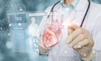
Nanotechnology has therapeutic potential in cardiology for arteriosclerosis as well as for hypoxic regions in the case of tissue ischaemia. The principle is the same as in imaging: ‘You need a suitable vector of nanoparticles, which must be coated in such a way (for instance with a receptor) that it reaches the desired target. The vector can also be equipped with drugs for drug delivery, or with stem cells for stem cell delivery,’ Prof. Pries points out.
Functional processes made visible
Through precise targeting of the stem cells the affected tissue regenerates far better after a heart attack compared to their injection into the bloodstream. In this case, the stem cells would travel to where it is attractive for them, rather than where they are actually needed. ‘Especially in cardiology, where the boundaries of myocardial tissue need to be reached, targeting is extremely important. This can, for instance, be achieved through anchorage to a changed endothelium. The vascular system is the best means of reaching all cells.’ Even though nanotechnology in medicine is still experimental rather than an everyday procedure in many areas and the effectiveness can only be ascertained at the end of the process, Prof. Pries sees considerable potential in this field, which requires further research. ‘Nano-imaging, as yet making up only a small part of functional imaging, has advanced into clinical practice the most, as functional contrast media make processes visible which previously were not visible at all or only visible via PET-CT. This delivers an entirely new quality of diagnosis,’ the professor points out, although also warning: ‘Nanotechnology has so far been viewed very positively. However, through uncontrolled use in everyday objects nanoparticles can also be detrimental to health. Although dosing for medicinal purposes has so far been considered safe.’
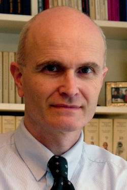
Professor Axel R Pries studied medicine at the University of Cologne and was habilitated by the Free University of Berlin (FU), where he began work as a research assistant in its Physiology Department in 1985. Ten years later he was an Associate Professor. In 1997, he became Senior Consultant at the Institute of Anaesthesiology, at the German Heart Centre in Berlin. Since 2001 he has headed the Physiology-CBF Department at Charité-Berlin, where he is also a Board Member of the Medical Faculty and Vice-Director of the Centre for Preclinical Medicine and the Centre for Cardiovascular Research at Charité.
25.08.2012