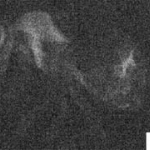
Thin client products aim to `unleash the potential of scanner technologies´
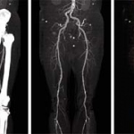
The Visage Thin Client product range on show at the ECR provides a fully-integrated system with advanced tools for 2-D, 3-D, and 4-D image review and interpretation, post-processing, data management, and image distribution.