High-Frequency Eyeball Sonography in the Differential Diagnosis of Papillae Changes
Since the beginning of the 90s, sonography has been a solid diagnostic pillar in ophthalmology centers, but it is also used by related medical disciplines, such as pediatrics, whose patients require ophthalmological treatment. For all age groups, the fundus of the eye, and especially the optic disk, is well-suited for high-frequency sonography with linear transducers. This type of examination ideally complements refraction study of the eye's fundus when suspicious changes in the papilla require further explanation. In such situations, the following differential diagnoses need to be considered: choked papilla from papilloedema, and optic nerve head drusen from pseudo-choked papilla and vascular occlusion.
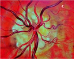
Fig. 1: c) ophthalmoscopic evidence of drusen of the optic nerve head
This article was first published in the VISIONS, issue 6/2004, a publication of Toshiba Medical Systems
13.08.2007
More on the subject:More on companies: