3D Vascular Imaging

Imaging of blood flow with colour Doppler sonography is an established method in prenatal medicine. Today, there are several techniques which offer colour visualisation whose quality is on a par with that of the B-mode. One example of such a technique is Advanced Dynamic Flow (ADF). Looking at two cases, we will describe the clinical use of ADF to visualise fetal vascular tumours. The quality of blood flow imaging with ADF is similar to that achieved with conventional colour Doppler. Since ADF uses a different frequency range than colour Doppler the colour is not superimposed on the B-mode. This avoids blooming artefacts and provides good delineation. Particularly, the visualisation of periphery and neighbouring vessels is improved with ADF. The insonation angle is now of minor importance. However, ADF is not able to depict turbulence.
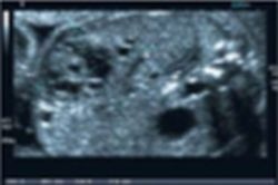
Fig. 1: Fetal giant haemangioendothelioma, initial diagnosis, B-mode

This article was first published in the VISIONS, issue 07/2005, a publication of Toshiba Medical Systems

23.07.2007

Related articles

Photo

Sponsored • Computed Tomography

Mobile Imaging Solutions: Clinical Confidence Anywhere

To bridge new equipment installations in radiology, temporary high workloads, or long-term additional capacity, Canon Medical provides mobile solutions according to customers’ needs and wishes.

Photo

Video • Redesign & refresh

Canon Medical updates Aplio ultrasound systems

Canon Medical announced the commercial launch of the Aplio i-series / Prism Edition, a complete redesign of its premium ultrasound series. In addition, Aplio a-series, Canon Medical’s…

Photo

Sponsored • All-in-one imaging trailer

Mobile solutions for the COVID-19 frontline

With public health issues continuing to make daily mainstream news headlines across the world, it is clear how much change the healthcare environment is going through. Not only are there existing…

Related products

Subscribe to Newsletter