Tumour Characterization Using Micro Flow Imaging

Due to echogenicity differences in comparison with the surrounding liver tissue conventional B-mode-imaging sonography permits the unambiguous classification of the frequently occurring typical liver cysts (criteria: round, echo-free, smoothly demarcated, with edge shadows and sound through transmission) and calcifications (highly reflective, acoustic shadows). The detection and characterization of liver tumours, however, continues to be a challenge to imaging, despite technical advances in sonography, computed tomography, and MRI.

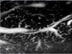
Rabbit liver as an example of the Micro Flow technique

This article was first published in the VISIONS, issue 07/2005, a publication of Toshiba Medical Systems

31.07.2007

Read all latest stories

Related articles

Photo

Sponsored • Computed Tomography

Mobile Imaging Solutions: Clinical Confidence Anywhere

To bridge new equipment installations in radiology, temporary high workloads, or long-term additional capacity, Canon Medical provides mobile solutions according to customers’ needs and wishes.

Photo

Video • Redesign & refresh

Canon Medical updates Aplio ultrasound systems

Canon Medical announced the commercial launch of the Aplio i-series / Prism Edition, a complete redesign of its premium ultrasound series. In addition, Aplio a-series, Canon Medical’s…

Photo

Sponsored • All-in-one imaging trailer

Mobile solutions for the COVID-19 frontline

With public health issues continuing to make daily mainstream news headlines across the world, it is clear how much change the healthcare environment is going through. Not only are there existing…

Related products

Subscribe to Newsletter