From head to toe
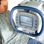
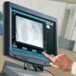
The new Philips 256-slice Brilliance iCT came in to use recently at the University Hospital in Ulm. The system produces quick, high-res scans with 80% less radiation.


The new Philips 256-slice Brilliance iCT came in to use recently at the University Hospital in Ulm. The system produces quick, high-res scans with 80% less radiation.
As the opening of the Beijing 2008 Olympic Games nears, the US Olympic Committee (USOC) and the General Electric Company (a Worldwide Partner of the Olympic Games) are running two research programmes aimed at demonstrating that health monitoring and early intervention leads to injury prevention and enhanced health and sports performances for athletes.
Before discussing a possible connection between gadolinium and nephrogenic systemic fibrosis (NSF), Dr Herborn offered a brief background:
From 2000 to 2007, Israeli hospitals have treated the victims of 148 terrorist attacks.

During ECR 2008, the Canada-based Acceleware Corporation, which develops acceleration solutions for high-performance computing, demonstrated its new AxRecon image reconstruction solution for medical imaging, security, and non-destructive testing.
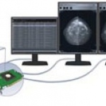
Combining the advantages of a traditional hospital light box with the features of computer workstations, BrainLAB has produced the Digital Lightbox - which is certainly unlike the usual light box.
Visage Imaging has launched its Cardiac Analysis software for the Visage CS Thin Client, which offers 'advanced visualisation and quantitative analysis for cardiac CT studies, such as calcium scoring, coronary artery analysis and left ventricle analysis,' the firm reports.

Olympus, which recently opened the global Research and Development Centre for Life Science in Munich, will invest around euro 15m in this project.
Erich Reinhardt, CEO of Siemens Healthcare, will resign effective April 30, in conjunction with compliance violations within Siemens' medical group. Reinhardt, 61, was not personally involved in the alleged corruption, according to Siemens' officials.
Hospitals concerned about the accuracy of utilization predictions of new diagnostic imaging equipment will be interested in pay-per-use financing that is being offered by global vendors. Eric-Jan Rutten, General Manager of Professional Healthcare Solutions of Philips Healthcare International in Eindhoven, Netherlands, described the approach that Philips takes with both large and very small…
A national electronic patient records (EPR) archive that is estimated to manage 550 pentabytes (PB) of data by 2025 is nearing completion in Finland. Its first component, an ePrescription system, is scheduled for implementation in June 2008. The patient record archive will be activated in February 2009, and DICOM diagnostic images will be added to it in the June 2009 time frame.
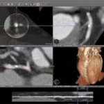
Encouraged by the success of the Dual-Source CT system Somatom Definition with two X-ray tubes that simultaneously generate different energies, Siemens Healthcare has already developed six specific dual energy applications. At the ECR 2008, the company presents four new applications that simplify the diagnosis of diseases of the heart, brain, lungs and extremity joints.
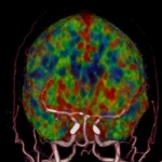
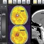
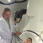
During a Toshiba press conference on Monday at ECR 2008, Prof P. Rogalla Chief Radiologist CT and Prof R. Klingebiehl, Department of Neuroradiology both of the Charité University Hospital, Berlin (Germany), reported about the new diagnostic opportunities the company's latest innovation — the Aquillion ONE - offers.
A 320-row CT scanner (Aquilion One, Toshiba Medical Systems Co., Tokyo, Japan) was installed for the first time in Europe, at the Charité University Hospital, Berlin, Germany, in November 2007. Its capability to cover the whole brain in a single rotation means this new type of scanner has the potential to impact strongly on the field of neuro-imaging.
'How do you treat the HIV-positive, diabetic, schizophrenic patient presenting with chest pain? By making the necessary information available for personalised medicine'
At this years ECR, Matrox, leading manufacturer of specialized graphics solutions for professional markets such as medical imaging, launched the Matrox Xenia Series controller boards. Supporting a wide range of displays and formats, each Xenia PCIe x16 single-slot board drives up to three high-resolution digital displays, minimizing time required to install, configure and deploy radiology and…
Magnetic resonance imaging is increasingly gaining importance as a second imaging process in prenatal diagnosis in addition to ultrasound examination, Dr. Daniela Prayer, a pediatric radiologist of the University Clinic for Radiological Diagnostics at the Vienna University Hospital, told reporters at a press conference held Friday.

Ziehm Imaging, provider of mobile X-ray based imaging systems (C-arms) and the French medical technology company Praxim announced a strategic partnership to utilize long established intraoperative 3D imaging technology for navigated surgery.
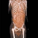
A new volumetric X-ray application, showcased at the European Congress of Radiology in Vienna, Austria, provides physicians with multiple high-resolution slice images of the human anatomy, including the chest, abdomen, extemities and spine.
Agfa HealthCare presents its entire Computed Radiography (CR) solutions range at ECR 2008 in Vienna. From desktop and compact solutions to groundbreaking Computed Radiography systems that fill the gap between CR and DR (Direct Radiography), the company is able to offer its customers the right solution for every facility of any size.
Bayer Schering Pharma presented the latest data from a health-economic evaluation of Primovist in the diagnosis of liver metastases. The results indicate that MRI using the liver-cell-specific contrast medium can lead to cost savings due to improved surgical treatment planning and less need for changes during operations.
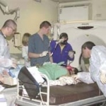
Over the last three decades CT has become a premier diagnostic tool for the evaluation of the acute patient. Over the past ten years in Israel, we have seen an overwhelming increase in the volume of CT examinations in the emergency department (ED).
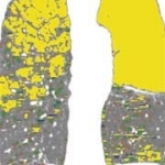
The Uliazpi Foundation in Spain, which studies and cares for severely mentally retarded patients, carried out an interesting study to identify bone mineral density values in a group of its patients, compare these with the general population and investigate the possible influence on these values on certain clinical variables and therapeutic regimens.
The Virtual Physiological Human By Hans-Ulrich Kauczor MD PhD, Director and Chairman of Radiology at Heidleburg University Clinic, and radiologists Frederik Giesel MD MBA and Hendrik von Tengg-Kobligk MD of the German Cancer Research Centre in Heidleburg, Germany.
Professor Rémy-Jardin MD PhD heads the Department of Radiology and is Chairman of the Department of Thoracic Imaging at the Calmette Hospital, University Centre of Lille. She is also Professor of Radiology in Lille University's Medical Faculty.