CT Fluoroscopy
In recent years, interventional procedures using realtime image control by means of Ultrasound have been increasingly integrated into the clinical routine as diagnostic and therapeutic procedures. However, a series of limitations in ultrasound result tram physical effects that interfere with the image. Combining fluoroscopy with a computed tomograph (CT) results in a procedure that enables realtime image control over the entire body with high geometric accuracy and for the most part without significant interfering artifacts, resulting in increased target accuracy, reduced intervention times and improved visualization of the biopsy needle. Depending on the procedure being used, higher radiation doses may occur than in conventional CT-supported interventions. Since the radiologist is present in the CT room during the intervention, he or she is exposed to additional radiation - a point that must be stressed because since 1994 over 65 cases of fluoroscopically induced radiation damage to the skin of patients and intervening radiologists have been reported and the number of unknown cases is probably much higher. Thus effective exposure reduction is extremely important for radiologists and patients alike and a number of options are examined in this study.
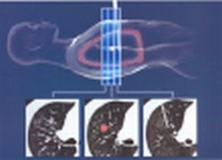
with three simultaneous slice images
This article was first published in the VISIONS, issue 4/2003, a publication of Toshiba Medical Systems
17.08.2007


