Contrast Harmonic Imaging (CHI)
Case 1 50-year-old male with a colorectal cancer.
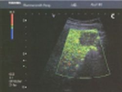
Greyscale imaging clearly shows the lesion, as an echogenic 3 cm mass (Fig.l a), which must be considered as suspicious for a metastasis from his colorectal cancer but benefit of the doubt must be given. Colour Doppler (Fig. 1b) is not helpful. At 33 seconds after SonoVue injection (early sinusoidal phase), Vascular Recognition Imaging (VRI) indicates a low vascular volume (Fig. 1c). This strongly confirms the diagnosis of a metastasis. Changing to Pulse Subtract Imaging on a second injection an additional lesion is clearly demonstrated high in the liver laterally (Fig. 1 d). This lesion was not apparent on the baseline B-mode image. Both lesions are confirmed on the CT scan (Fig. 1 e,f).
This article was first published in the VISIONS, issue 4/2003, a publication of Toshiba Medical Systems
20.08.2007
More on the subject:More on companies: