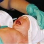
Paediatrics

Photo

FUNDRAISING

When the Oxford Radclife Hospitals NHS Trust invested £109 million in its new Oxford Children's Hospital, funding for certain special embellishments could not be contemplated. Thus a £15 million Campaign was launched to enable the hospital to be built and equipped far beyond the NHS standard. £13.8 million of that target has so far been received. Who raised that astonishing sum? Its…

Photo

NEW VISIBILITY FOR NECKER-ENFANTS MALADES in Paris

In the heart of Paris, the Necker children's hospital, the oldest in the world dedicated to paediatrics, was suffocating inside a completely inward-looking enclave. The challenge was to achieve a vast upheaval to overcome the hospital's lack of relationship with its surroundings and open it onto the city, while also carrying out an in-depth metamorphosis to adapt it to new requirements.

Photo

First Czech "test-tube" baby celebrated 25th birthday

The first Czech test-tube baby was born, woman in childbed delivered smoothly on 4th November 1982, and healthy baby-boy was 51 cm tall, and weighed 3.65 kg. A medical team from Moravian town of Brno led by embryologist Milan Dvorak and gynecologist Ladislav Pilka was behind all this happy-ended artificial fertilization quarter a century ago.

Photo

Pulse oximetry - screening tool in newborn babies

A research carried out at six hospitals in the West Midlands, UK, is evaluating the use of pulse oximetry as a screening tool for congenital heart disease in newborn babies. Approximately three percent of infant deaths are caused by these significant heart defects and at the moment only less than half of the affected babies are identified by clinical examination, the current screening technique.

Photo

Close to baby

Every young mother wants to be as close as possible to her newborn baby. The "Leipiziger Wanne", the "Leipzig tub", developed by German engineering company NEL, can fulfil this wish even more easily: The c-shaped bed consists of a transparent tub which can be moved across the mother's bed allowing her eye contact with the baby at any time.

Photo

Paediatric surgery: a major challenge

UK — A report on the care of young surgical patients has been launched by The Children's Surgical Forum, a body of representatives from the medical royal colleges, surgical specialist associations, Department of Health, Royal College of Nursing and the Royal College of Surgeons Patient Liaison Group.

Photo

Sonographic Indications of Fetal Chromosome Abnormalities

"Genetic ultrasound" is a refined technique in prenatal diagnostics. It aims to estimate as accurately as possible and non-invasively, the individual risk of a fetal chromosome abnormality. In conventional prenatal sonography (malformation screening) conspicuous fetal malformations are searched for selectively; naturally chromosomal anomalies of the fetus cannot be detected in this…

Photo

New Image Guided Therapy Suites at Sick Kids

In May 2001, the world's first CT-XR hybrid suite for image guided therapy in paediatrics was put into operation. The new Centre for Image Guided Therapy (IGT) at The Hospital for Sick Children allows surgeons and interventional radiologists to do something that's never been done before - work side-by-side when diagnosing and treating children. A total of 4 interventional suites were equipped…

Photo

Looking at a New Generation

Most babies are born healthy, but there is still a small risk for all women to give birth to a baby with some type of anomaly. Ultrasound has proven to be one of the most reliable procedures to prenatally diagnose some of these conditions. Ultrasound can be used during the entire pregnancy, but in the first trimester ultrasound examinations are, as a rule, used to exactly calculate the pregnancy,…

Photo

Advancing Pediatric Imaging with 16-Slice CT

Pediatric imaging presents many unique and difficult challenges to the radiologist. Correct and rapid diagnosis is extremely important in pediatric imaging and to that end we constantly strive for tests that offer more information, accuracy and versatility. However, it is also our duty to provide tests that are safe and minimise exposure to ionizing radiation. In recent months there has been…

Photo

High-Frequency Eyeball Sonography in the Differential Diagnosis of Papillae Changes

Since the beginning of the 90s, sonography has been a solid diagnostic pillar in ophthalmology centers, but it is also used by related medical disciplines, such as pediatrics, whose patients require ophthalmological treatment. For all age groups, the fundus of the eye, and especially the optic disk, is well-suited for high-frequency sonography with linear transducers. This type of examination…

Photo

Screening for Chromosomal Defects

Chromosomal abnormalities are major causes of perinatal death and childhood handicap. Consequently, the detection of chromosomal disorders constitutes the most frequent indication for invasive prenatal diagnosis. However, invasive testing, by amniocentesis, chorionicvillus sampling (CVS) or cordocentesis, is associated with a risk of miscarriage of about 1% and therefore these tests are carried…

Photo

The Non-Surgical Closure of Congenital Heart Defects

What is an “open foramen ovale”? The open foramen ovale medical abbreviation PFO for “patent foramen ovale”) is a usually small, only a few millimeter thick, membrane-covered slit in the septum between the atria of the heart. It is a remnant from human embryonic development that usually closes during the newborn period; however, in 10-25% of newborns a small opening remains that generally…

Photo

Robust CT Protocols for Pediatric Cardiovascular Examinations

Pediatric cardiovascular disorders are extremely complex in nature and present with a widely variable array of abnormal anatomy. Imaging these disorders presents a unique set of challenges for the cardiovascular team. Complex anatomical variations require detailed volumetric image interpretation with high spatial and temporal resolution. In addition, the patients are invariably young. They may…

Photo

Tissue Doppler Imaging of the Fetal Heart

In the last 25 years, fetal echocardiography has become established as an integral component of prenatal medicine. Structural heart defects are found in approx. 0.8% of all foetuses. They are consequently comparatively frequent and head the list of isolated fetal organ malformations. Routine screening of the four-chamber view and the outflow tract allows the experienced investigator to detect…

24 show more articles
Subscribe to Newsletter