The advantages of ultrasound for paediatric diagnosis
A presentation for the 31st tri-national DEGUM, ÖGUM, and SGUM conference in Leipzig, Germany, by Axel Feldkamp MD (below), Chairman of the DEGUM paediatrics division, and Director of the paediatrics department at Duisburg Hospital.
Ultrasound is the most important imaging technique in paediatrics – more so than in any other branch of medicine. This is attributable to a number of factors. X-ray exposure is a more significant risk factor for paediatric patients than for adults by virtue of the cumulative nature of X-ray dosages, thus making paediatric patients more likely to receive a higher dose of radiation. Moreover, some types of immature tissue are more susceptible to radiation. Thus it is essential to avoid the use of X-rays in children and young adults.
Ultrasound scanners have now evolved to the point where the image quality they deliver obviates the need to use any other imaging technology. Various types of heart catheterisation can now be realised using ultrasound in lieu of radiography. Elaborate high-radiation imaging procedures for the urinary tract are increasingly replaced by the use of ultrasound contrast media. Complex imaging of skull pathology in paediatric patients can now be realised using ultrasound rather than radiography.
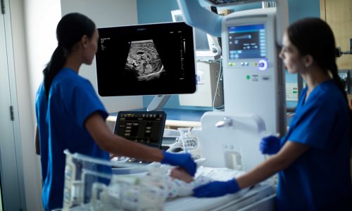
Another advantage of ultrasound examinations lies in their ready availability. In neonatology in particular, examinations of neonates in incubators are indispensable, since moving the child from one place to another, wrapped in a textile, would constitute an extreme risk. In addition, thanks to the progressive miniaturisation of ultrasound scanners, paediatric ultrasound examinations can be realised pretty much anywhere and at any time.
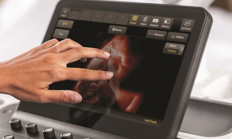
High resolution ultrasound scanner heads (ideal for neonatology by virtue of their relatively shallow penetration depth) often produce image quality that is unmatched by any other method. In some cases, only ultrasound can detect congenital heart defects, brain morphology defects, cranial bleeding, and haemodynamic complications. Although MRI provides outstanding image quality in infants, the examinations are lengthy and an infant must be either anaesthetised or heavily sedated.
Ultrasound is taught in paediatric training programs owing to the extreme usefulness of the technique. Consequently, ultrasound is widely available and can be used for screening purposes. For example, the current practice of doing a hip screening for all newborns has reduced to a minimum the number of hip defects requiring surgery.
Thus ultrasound can be regarded as the paediatric imaging technique of choice for the following reasons:
- the child is not exposed to radiation
- ultrasound devices can be deployed just about anywhere
- ultrasound image quality constantly improves, thanks to the use of high-resolution transducers.
14.11.2007