More stable contrast media expand the ultrasound spectrum
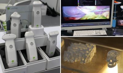
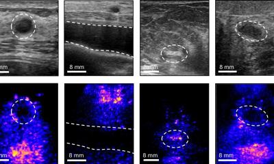
Due to significant developments in ultrasound contrast media and equipment technology even the finest blood vessels can be visible through contrast-enhanced sonography.
In many areas, e.g. tumour or infarction detection, image quality today provides as much informative value as X-rays or CT scans. Since 1986, Professor Kurt A Jäger, Senior Consultant at the Department for Angiology at the University Hospital Basel, Switzerland, has been working with and researching contrast-enhanced sonography. He is also vice president of the International Contrast Ultrasound Society (ICUS) founded in September 2008.
‘Significant improvements in ultrasound technology and the consistency of contrast media have let this procedure mature,’ he says. ‘The new high-end equipment not only has improved colour but also much better image quality and resolution. So, in many areas there’s no longer even a need for contrast media. However, for certain indications it’s possible to gain a lot of information in a relatively economical way by using modern contrast media, i.e. everywhere where we need to know more about the organs’ functional efficiency. Previously, air bubbles in the contrast media were too large to enter arterial circulation via the lungs. However, helped by the second generation of contrast media, with smaller gas bubbles it became possible in the late ‘90s to achieve a much improved view of circulatory disorders of the legs, neck and head, as well as abdomen, i.e. bowel or kidneys. The new generation of contrast media and sonography equipment has now made it possible to benefit from the full potential of the procedure and to show not only macrocirculation but also blood flow everywhere in the body on a micro-circulatory level.
‘Compared with CT, there are decisive advantages. Using ultrasound you can capture very fast sequences continuously, whereas CT only captures individual sequences, possibly missing significant information if you don’t detect in the right time phase. On the other hand, ultrasound contrast media remain in the vessels, whereas CT contrast media diffuse out of the tissue after a short time, so they deliver only incomplete information about the microcirculation.’
Speaking of the extent that the development of contrast media and the technology have been closely linked, Prof Jäger points to what he considers one big step forward: ‘Nonlinear vibration of the gas-filled bubbles, which are administered as contrast media, can also be detected with this equipment. The air bubbles start to oscillate in the ultrasound field and it’s only that which makes the blood flow in the tissue visible on the screen. The effects of the contrast media can be even better shown with the new equipment.’
Worldwide controversies about contrast media have repeatedly delayed this procedure’s implementation, largely because, in very rare cases (0.001%), patients with severe heart disease can experience problems with contrast media administration. However, in 2004 product warnings from the European Pharmacological Control Office, and in 2007 from the US Food and Drug Administration, led to discontinuation of contrast media use in the respective countries. Contrast media licensing for radiological and medical diagnostics is very different in various countries. ‘Europe is more advanced than the USA; we have been working towards standardisation for quite some time,’ the professor points out. ‘If we now begin to exchange our knowledge internationally then we will progress infinitely quicker in offering patients a safe procedure that does not put any strain on them.’
As for the aims of the new International Contrast Ultrasound Society (ICUS), he explains there are various doctors using contrast media and they will be encouraged to combine their know-how. ‘Our objectives are standardisation, education and training. Often the various disciplines know nothing about common problems or each other’s expertise.’
21.11.2008