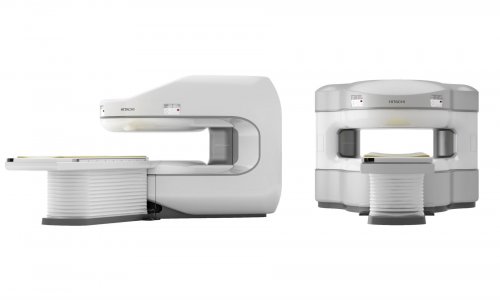
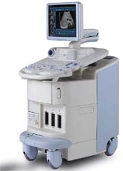
HI VISION 900
Hitachi has developed a new abdominal sonography system, based on a fully digital platform with a unique high-performance computer and an innovative octal parallel wideband beamformer, the firm reports.
‘The system offers maximum resolution with optimum penetration depths in combination with the Gig Sampling technology with 12-bit A/D conversion and the multi-frequency wideband imaging for all clinical situations and, with its dynamic bandwidth of over 150 dB, it offers accurate resolution of the finest organ structures even in the native B-image. The current range of more than 45 different types of transducers offers not only an unrestricted range of applications but it is also compatible with all other Hitachi ultrasound systems.’
Accurate display of smallest lesions
With its comprehensive standard equipment the system has technological features that guarantee incredibly accurate imaging, Hitachi points out. ‘The 2nd generation SonoMR technology, for example, significantly reduces image artefacts while increasing the detail and contrast resolution, which allows improved differentiation of subtle lesions and clear display of tissue margins and transition zones.’
Many display types
‘Because a different system mode enables the best diagnoses for every application,
the HI VISION 900 offers complete flexibility, allowing the user to select 2-D imaging with optimum detail and contrast resolution, M-mode and Colour-M-Mode display with maximum time resolution and digital, controllable PW/CW Doppler, Colour Flow Imaging in real-time and with true-colour processing, Colour Flow Angio display with maximum sensitivity and bidirectional Colour Flow Angio mode with intensity display while simultaneously displaying the direction of flow.’
Real-time Tissue Elastography (HI-RTE)
This measures and displays tissue elasticity in real-time. ‘Tissue is compressed slightly with the standard transducers and the information is colour-coded and superimposed over the native B-image. Different colour scales allows a transparency display. The native B-image and tissue elasticity can also be displayed in parallel in the dual image. To ensure that an organ perfusion can be displayed down to the finest details in combination with ultrasound contrast agents and lesions can be correctly visualised and evaluated, dynamic Contrast Harmonic Imaging uses the Wideband Pulsed Inversion technique (WPI), which enables localisation of contrast agents and eliminates native and harmonic tissue signals. Different modes are available in this procedure in low Mi and in high Mi scanning.
Display in 16 million colours
The 17-inch computer graphics display with maximum resolution, flicker-free
image frequency, true-colour display in 16 million colours and digital RGB reproduction of even video in-put signals guarantees stress-free examinations. A remote control and voice control are also available for operation in a sterile environment in restricted spaces.
19.11.2008