Elastoscan in mammary sonography
By V. Duda, of the Senological Diagnostics Department, Giessen, and University Hospital Marburg
If, in the early days of mammary sonography, it was revealed to be helpful to render tactual findings in a visible manner (sonic palpation), then now is the time to palpate clinically occult findings in a new manner (Elastoscan).
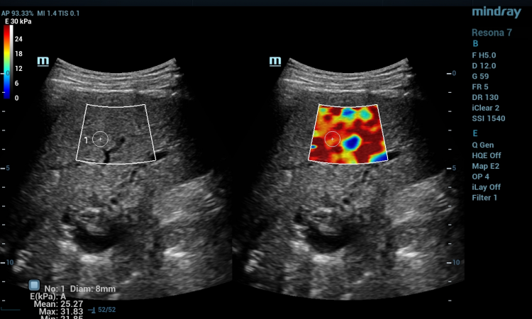
The use of the ‘strain effect’, i.e. checking to what extent reflectors can be separated from one another under compression, provides important additional information on tissue conditions in a region of interest not to be further differentiated by the B-mode image.
The complementing of conventional B-mode sonography by elastography represents, by contrast to Doppler sonography and 3-D sonography, a much less time-consuming new approach that is available without any additional preparations.
For more enhanced requirements made of sonography in early detection of breast cancer it is increasingly important to also unerringly address such focal points as would scarcely have been remarked upon without mammographic detection with ultrasonics. In addition, these focal points are, in part, only rudimentarily to be differentiated from the small intra-mammary haematomas frequently occurring in pre-operative interventions.
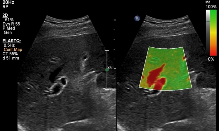
Similar indications for elastography arise during ‘second look’ sonography during the search for correlates of mamma-MRT findings and in improved targeting for the execution of interventional measures (punch biopsy) on small or difficult to delineate focal findings.
Additionally, still under scientific evaluation are questions of detail definition of therapy monitorings, whether in the case of pre-operative chemotherapy, pre-operative predictions on size and the supplemental sonography of mammary excidates.
Gathered previous clinical experience shows that elastography does not, in fact, actually replace mammary sonography in histological clarification but, particularly in the case of small or more deeply situated findings, delivers unambiguous and clearly reproducible images. Hence, clearly targeted work becomes a possibility and a further, promising facet provides an enhancement to sonography, a facet the deployment possibilities of which have scarcely even been embarked upon.
* Elastoscan is a trademark of Medison
19.11.2008