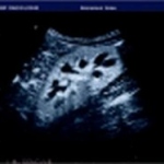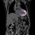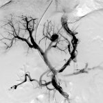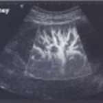
Aplio - Dynamic and Advanced Dynamic Flow
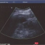
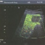
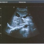
There are a number of problems in contrast imaging using LevovistTM contrast agent. In Doppler mode, the problems are poor resolution and large areas of blooming. In 2nd harmonic imaging and pulse inversion imaging, there are problems related to tissue harmonic imaging (THI) because Levovist requires a high mechanical index (MI). THI increases under high-MI conditions, and THI interferes with…