Article • Cardiovascular care at a continental scale
The case for imaging in Europe’s Safe Hearts Plan
Cardiovascular disease remains the leading cause of death across the EU, cutting short the lives of millions of Europeans every year, and placing an enormous strain on health systems and economies alike. Yet prevention and early intervention can have a major impact – and this is precisely the ambition of the EU Safe Hearts Plan. At a high-level event at the European Parliament in Brussels in July, policymakers, clinicians, patients, and industry came together to explore how diagnostic imaging can contribute to this goal.
Report: Wolfgang Behrends
Image source: © ImageFlow / Luke – stock.adobe.com
© European Union - Source: EP; photographer: Philippe Buissin
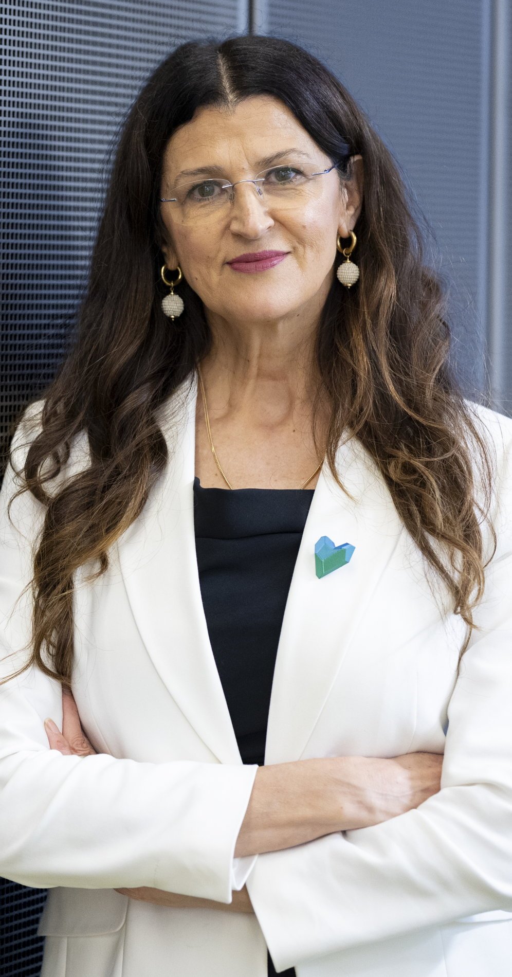
The event, hosted by MEP Romana Jerković, Vice-Chair of the European Parliament's Committee on Public Health and Chair of the MEP Group on Cardiovascular Health, and co-organised by the ESR and EANM – who launched dedicated policy briefings for the occasion – opened with a clear statement of intent. ‘Europe must go beyond a model centred primarily on treating cardiovascular disease after a clinical event,’ said Jerković, who served as rapporteur for the European Parliament's own-initiative report on EU cardiovascular health strategy. ‘We need a health policy that places greater emphasis on prevention, on early detection, timely diagnosis, and personalised care.’
Adopted by the European Commission in 2025, the Safe Hearts Plan is Europe's first comprehensive strategy for cardiovascular health, built around three pillars – prevention, early detection and screening, and treatment and care.1 Medical imaging, Jerković argued, is central to all three. ‘Advanced imaging is a key component of the cardiovascular care transformation, but no single profession and no single institution can deliver this transformation alone.’
Vytenis Andriukaitis, MEP, a cardiac surgeon by training, placed this cardiovascular strategy within the broader goal already being pursued through the European Health Union – a policy framework launched during the Covid-19 pandemic to strengthen the EU's fragmented health crisis management. Building upon the recently established European Health Data Space (EHDS), the Safe Hearts Plan could become the foundation for a continent-wide imaging network. Used to exchange data, harmonise diagnostic standards, and reach patients from Helsinki to Lisbon, this interconnectivity would greatly strengthen the European Health Union as a whole, he argued.
The radiological perspective

Image source: Radboud UMC
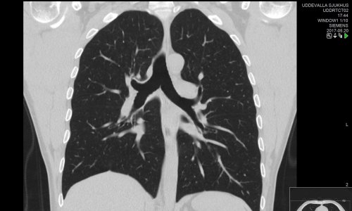
‘The Safe Hearts Plan marks a historic shift from reactive treatment to proactive prevention,’ said Professor Matthias Prokop, Head of the Department of Medical Imaging at Radboud University Medical Center in Nijmegen, the Netherlands, and ESR President – and imaging, he argued, is indispensable to making that shift a reality. For him, the key challenge lies in finding ways to maximise patient benefit by cleverly using existing diagnostic resources. For instance, cardiac MRI scans can also be leveraged to detect cancers – and vice versa, CT images taken for lung cancer screening may reveal coronary calcium, a strong predictor of adverse cardiac outcomes.2 ‘It is important that we use the expertise that we have in radiology to get these synergies,’ Prokop said.
This ties in with Europe's Beating Cancer Plan, another pillar of the European Health Union, which has driven a significant expansion of lung cancer screening programmes across the continent. The same data, Prokop added, can also be used to avoid unnecessary treatments – for example, withholding statins from a patient with a low coronary calcium score. ‘It is existing data that can help us make our population healthier with a minimum of extra effort.’ The infrastructure, in other words, is already in place – what is needed is the will to use it systematically.
Image source: EANM
Professor Anna Erba, Associated Professor at the Department of Medicine and Surgery at the University of Milan Bicocca, Italy, and EANM President, cited practical clinical examples to underline how imaging is improving diagnostic certainty, supporting personalised care pathways, and contributing to a more efficient and sustainable healthcare system. ‘To put it simply: images become numbers, those numbers become knowledge, and that knowledge becomes better care and better value for our health systems.’ She called on delegates to view radiological and nuclear medicine imaging not as alternatives but as complementary approaches. ‘In this multidisciplinary framework, collaboration is key,’ Prof. Erba stressed, ‘but we also have to take care of our workforce, because without properly trained imaging professionals, innovation does not reach the patient.’
Dr Rodrigo Salgado, radiologist at Antwerp University Hospital and Holy Heart Hospital Lier, Belgium, and President of the European Society of Cardiovascular Radiology, grounded the argument in clinical evidence. The SCOT-HEART trial,3 he noted, has demonstrated that adding cardiac CT to standard care in patients with stable chest pain improves diagnostic certainty and patient outcomes at ten years – using nothing more than existing scanners, standard software, and basic cardiac radiology knowledge.4 ‘The infrastructure is already in place,’ he said. ‘Nothing more is needed than just to implement it’ – a conclusion now reflected in the European Society of Cardiology's recent guidelines, which establish cardiac CT not as a luxury but as a clinical necessity.5
Nuclear medicine: from structure to function
Professor Lars Christian Gormsen, clinical professor of nuclear medicine at Aarhus University, Denmark, underlined that anatomical and functional imaging are most powerful in combination: where radiology establishes the structural picture, nuclear medicine techniques such as PET can determine whether a stenosis is truly causing ischaemia, whether damaged myocardium is still viable, and whether a treatment is working – serving, as he put it, as ‘another gatekeeper before we send the patient to invasive procedures.’
Professor Riemer Slart extended this argument to a condition that remains largely invisible to conventional diagnostics: coronary microvascular dysfunction, which disproportionately affects women and frequently presents with typical chest pain despite normal stress tests and CT angiography. Functional PET imaging, he demonstrated, can identify these patients accurately – addressing an important gap in cardiovascular care and supporting more equitable diagnosis and treatment.
Recommended article
Article • Expert perspectives from ESC 2025
Faster, smarter, deeper: how new technologies redefine cardiac imaging
New cardiac imaging techniques continue to uncover the secrets of the heart for cardiologists who know how to use them. At the ESC 2025 Congress in Madrid, four experts explored cutting-edge developments across different modalities. These novel approaches provide a promising glimpse of what the next generation of cardiac imaging is capable of.
The future of cardiovascular imaging: three transformations
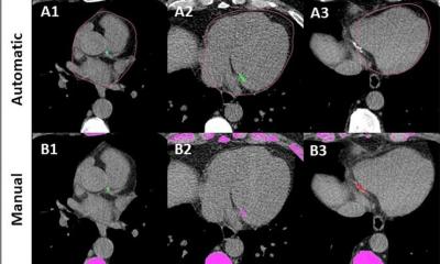
Looking further ahead, Professor Marco Francone, Chairman of Radiology at Sapienza University of Rome, Italy, outlined three major transformations in cardiovascular imaging that are already under way – and that align directly with the ambitions of the Safe Hearts Plan. The first is a shift from morphology to biology: images now reveal not only the presence of a plaque but its fatty composition, the inflammatory state of the surrounding pericoronary tissue, and the degree of downstream perfusion impairment – information that determines myocardial injury and infarction risk far more precisely than anatomy alone.
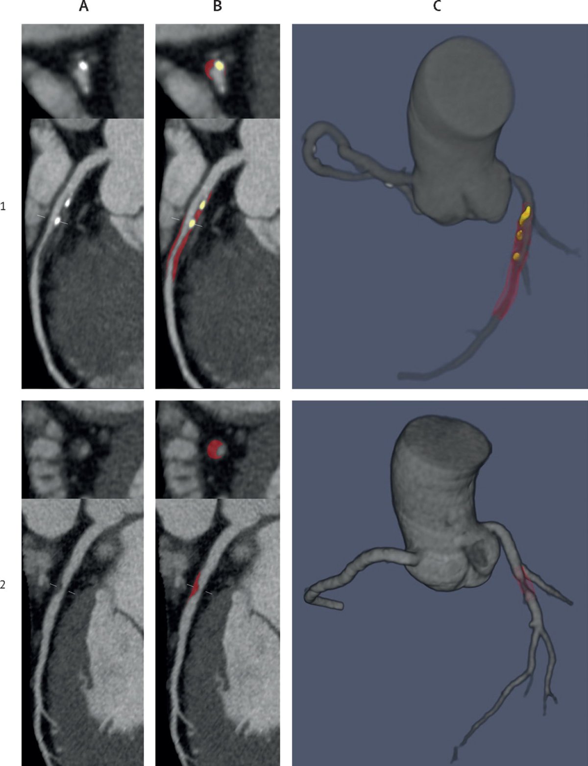
Image source: Lin A, Manral N, McElhinney P et al., Lancet Digital Health 2022 (CC BY-NC-ND 4.0)
The second transformation is from standardised protocols to personalised care. ‘Precision care means treating the right patient with the right approach – and imaging is driving this,’ Francone said, noting that AI and computational fluid dynamics now allow clinicians to quantify plaque burden, map calcification distribution, and model coronary flow in individual patients – shifting imaging from a descriptive tool into a guide for treatment decisions.
The third – and most ambitious – transformation is a move from individual to population care. With CT volumes in Europe having increased 4.5-fold over the past decade, Francone argued that the continent is sitting on a largely untapped resource. ‘We should exploit the millions of scans done yearly in Europe for better health,’ he said – building a European imaging network and biobank, with secure data sharing and AI-based biomarkers and prediction models, to extend the benefits of precision imaging to entire populations. A large study published in The Lancet has already demonstrated that AI-derived plaque volume measurements predict myocardial infarction risk with clinically meaningful accuracy,6 pointing the way towards a future in which imaging does not merely diagnose disease but anticipates it.
From Brussels to the bedside: policy and implementation
The panel discussion that closed the session turned from clinical evidence to the practical conditions for implementation. Professor Evis Sala, Minister of Health of Albania and a trained radiologist, spoke from the dual vantage point of imager and policymaker. ‘As imagers, we are transversal – we see the whole pathway,’ she said, pointing out the importance of a well-organised, orchestrated pathway with imaging as a key component for patient outcomes.
We cannot think about imaging without also tying it in with artificial intelligence – and when we are thinking of AI, we are thinking of quality datasets, structured reporting, and harmonised imaging protocols across Europe
Isabelle Baustert Chrysanthou
Isabelle Baustert Chrysanthou, Health Attaché at the Permanent Representation of the Republic of Cyprus to the EU, pointed to the conditions that will determine whether the plan's ambitions translate into practice. ‘We cannot think about imaging without also tying it in with artificial intelligence – and when we are thinking of AI, we are thinking of quality datasets, structured reporting, and harmonised imaging protocols across Europe,’ she said. Yet she also acknowledged the structural constraints: health remains a national competence, and the discrepancies between member states mean that political will at European level must be matched by concrete support on the ground. ‘Countries will need support to be able to do this together.’
Viktoriia Shportiuk, Co-Chair of the COCIR Cardiovascular Disease Task Force, brought the industry perspective. While welcoming the Safe Hearts Plan's focus on prevention, she cautioned that prevention alone is insufficient – proper referral pathways and access to confirmatory diagnostics are equally essential. She also underlined the importance of adequate funding – citing the European Cancer Plan as evidence that without financial support, equitable care across all member states remains out of reach – and called for a regulatory environment that allows industry to continue launching innovation in Europe first, while maintaining patient and practitioner safety.
Closing the session, MEP Jerković reiterated the speakers’ conviction that ‘advanced imaging is a cornerstone of modern medicine, which enables earlier detection, precise diagnosis, better prediction, and more personalised treatment – contributing not only to better outcomes, but also to a more resilient and more sustainable health system across the EU’.
References:
- European Commission: Cardiovascular health – the Safe Hearts Plan; https://health.ec.europa.eu/non-communicable-diseases/cardiovascular-health_en#eu-cardiovascular-health-plan-the-safe-hearts-plan
- Chiles C, Duan F, Gladish GW, et al. Association of Coronary Artery Calcification and Mortality in the National Lung Screening Trial: A Comparison of Three Scoring Methods. Radiology. 2015;276(1):82–90. doi:10.1148/radiol.15142062
- SCOT-HEART investigators. CT coronary angiography in patients with suspected angina due to coronary heart disease (SCOT-HEART): an open-label, parallel-group, multicentre trial. Lancet. 2015;385(9985):2383–2391. doi:10.1016/S0140-6736(15)60291-4
- Williams MC, Wereski R, Tuck C, et al. Coronary CT angiography-guided management of patients with stable chest pain: 10-year outcomes from the SCOT-HEART randomised controlled trial in Scotland. Lancet. 2025;405(10475):329–337. doi:10.1016/S0140-6736(24)02679-5
- Vrints C, Andreotti F, Koskinas KC, et al. 2024 ESC Guidelines for the management of chronic coronary syndromes. Eur Heart J. 2024;45(36):3415–3537. doi:10.1093/eurheartj/ehae177
- Lin A, Manral N, McElhinney P, et al. Deep learning-enabled coronary CT angiography for plaque and stenosis quantification and cardiac risk prediction: an international multicentre study. Lancet Digit Health. 2022;4(4):e256–e265. doi:10.1016/S2589-7500(22)00022-X
28.08.2026