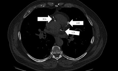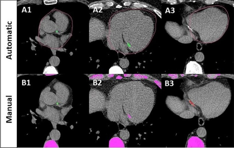
Image source: Hamelink I, Nie Z, Severijn TEJ et al., European Journal of Radiology 2025 (CC BY 4.0)
Article • Opportunistic screening
AI tools extract hidden health insights from routine chest imaging
Artificial intelligence is enabling radiologists to extract valuable diagnostic information from routine chest imaging – identifying patients at risk for osteoporosis and cardiovascular disease without additional scans. At RSNA 2025, researchers presented two AI-powered tools that transform standard chest X-rays and CT scans into opportunistic screening devices.
Special report: Cynthia E. Keen
An estimated 500 million people worldwide have osteoporosis or its precursor, osteopenia – yet most remain unaware of their condition.1,2 Because bone mass reduction is symptomless, the International Osteoporosis Foundation reports that up to 80% of people who suffer a fragility fracture will never be diagnosed or treated for the underlying disease.3
The gold standard for identifying osteoporosis is dual-energy X-ray absorptiometry (DEXA), which measures bone mineral density at the spine and femur. However, barriers to DEXA include limited scanner availability in underserved areas, demand exceeding capacity, a shortage of trained technologists, and cost.

Image source: Asan Medical Center
The chest X-ray is the most common imaging examination ordered by physicians, accounting for approximately 45% of all radiographic exams performed. In some countries, they represent up to 20% of all imaging.4 The ability to identify high-risk patients using a posteroanterior view chest X-ray could dramatically reduce the risk of future bone fractures.
At RSNA 2025, Namkug Kim, PhD, Professor at the University of Ulsan College of Medicine, Asan Medical Center in Seoul, South Korea, reported that a validation test of 77,677 chest X-rays from 44,773 patients – who also had a DEXA scan within six months – showed that the AI tool achieved 90% sensitivity and 81% specificity.1,5
The AI tool is sold under the name Osteo Signal by medical AI software developer Promedius. It has been cleared for commercial clinical use in Korea, Vietnam, and Indonesia. The company has applied for CE Mark and FDA clearance, with the hope for approval in late 2026.
AI turns chest X-rays into bone health screening
An AI-powered diagnostic support tool that analyses chest X-ray images for bone mass reduction risk may change this situation. The tool uses deep learning algorithms to identify radiographic patterns associated with osteoporosis risk and automatically generates a numeric score that is seamlessly added to the patient's radiology records.
Cardiovascular risk from routine chest CT
Researchers in Azerbaijan have developed an AI-powered quantification tool for cardiovascular risk stratification equivalent to dedicated cardiac CT. When a patient undergoes a standard non-cardiac chest CT examination, the screening tool calculates a coronary artery calcification (CAC) score automatically and incorporates this information smoothly into current radiology processes.
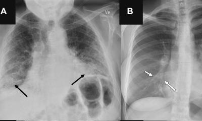
Photo courtesy of Dr. Elshan Abdullayev
In his RSNA 2025 presentation, Elshan Abdullayev, MD, Chief of Radiology at Saf Hospital in Baku, explained that the AI tool is not a replacement for a cardiac CT examination. 'Cardiac CT exams provide excellent risk stratification through calcium scoring, but a patient must be selected for this imaging procedure. Yet thousands of patients undergo routine non-cardiac CT exams performed daily which may contain visible coronary calcification that goes unreported. If identified and reported, these could provide life-saving cardiovascular risk information without additional radiation exposure or procedural burden.'
Reclassifying patient risk
In a retrospective study of 8,547 routine chest CT examinations from six academic centres to validate the screening tool, 2,847 patients with previously unclassified risk were identified and reclassified to higher categories. Of the total, 24.3% had their risk reclassified and 18.2% received interventions.
Dr. Abdullayev reported that the AI tool achieved excellent diagnostic accuracy for its automated CAC scoring across all severity categories, with a sensitivity of 94.8% and a specificity of 97.2%. The statistical analysis process, which also included survival analysis, took 12 seconds to perform.6
'Early identification of high-risk asymptomatic patients could represent a paradigm shift toward proactive population health management using existing imaging infrastructure,' Dr. Abdullayev emphasised. 'Early intervention enables timely initiation of evidence-based preventive therapies.'
Additionally, resources are optimised. The tool provides value from existing imaging infrastructure without significant modifications to the radiology department workflow. It rules out patients who do not have CAC risk and identifies at-risk patients who may be accidentally overlooked.
Dr. Abdullayev subsequently advised Healthcare in Europe that the AI quantification tool is in the research and validation stage. When it is ready for commercialisation, the tool's developers hope to partner with established medical imaging or AI companies that share a commitment to evidence-based, patient-centred innovation.
Profiles:
Namkug Kim, PhD, is a full Professor at the University of Ulsan College of Medicine, Asan Medical Center in Seoul, South Korea. He is co-founder of Promedius, a Seoul-headquartered company focused on aging and metabolic diseases. His research focuses on translational medicine, encompassing deep learning, 3D printing, computer-aided diagnosis and surgery, and medical image processing.
Elshan Abdullayev, MD, is Chief of Radiology at Saf Hospital in Baku, Azerbaijan, and Head of the Radiology Department at Misrata Heart and Cardiovascular Center in Misrata, Libya. He specialises in diagnostic radiology, with a particular focus on cardiovascular and thoracic imaging, MRI, CT, and the clinical integration of AI in medical imaging.
References:
- Kim M, Kim M, Kim N. “Opportunistic bone health screening: deep learning analysis of chest radiographs for osteoporosis and osteopenia detection.” Abstract T7-STCE2. RSNA 2025.
- 360 ResearchReports. “DEXA Bone Densitometers Market Size, Share, Growth, and Industry Analysis, By Type (Axial Bone Densitometer, Peripheral Bone Densitometer), By Application (Hospitals & Clinics, Universities and Research Institutions, Others): Regional Insights and Forecast to 2034. “Updated 16 February 2026. https://www.360researchreports.com/market-reports/dexa-bone-densitometers-market-211843. Accessed 26 February 2026.
- International Osteoporosis Foundation. “Osteoporosis: a global health crisis we can no longer ignore.” 20 October 2025. https://www.osteoporosis.foundation/news/osteoporosis-global-health-crisis-we-can-no-longer-ignore-20251020-1320. Accessed 26 February 2026.
- Radiopaedia. “Chest radiograph”. Revised 25 December 2025 by Stefan Tigges. https://radiopaedia.org/articles/chest-radiograph. Accessed 26 February 2026.
- Koo B, Roh Y, Shin G, et al. “Performance evaluation of deep learning-based osteoporosis diagnostic models with conventional chest x-ray in a clinical cohort.” J Thorac Dis 2025; 17(11): 10127-10137.
- Abdullayev E. “Predicting cardiovascular risk assessment from routine chest CT AI-driven coronary artery calcium scoring and aortic valve analysis.” Abstract S2-STCE1-1. RSNA 2025.
15.05.2026