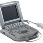
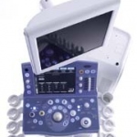
Photo

Selenium-Based Flat Panel X-ray Detector for Digital Fluoroscopy and Radiography

We have succeeded in developing the first selenium-based flat panel X-ray detector for digital fluoroscopy and radiography. This flat panel X-ray detector efficiently captures X-rays that have passed through the patient's body and converts them directly to digital dynamic and static images. It has been confirmed that images with high spatial resolution and contrast can be obtained. The detector…

Photo

Enviromental Protection in medical technology

For the production of medical technology products more and more companies take enviromental protection and energy efficiency into consideration. Siemens Medical Solutions e.g. ensures with a holistic concept that the products not only match high ecological demands during production, but also in operation and beyond shutdown.

Photo

Endoscopic Ultrasound Transducer with FNA Biopsy Capability

Endoscopic Ultrasound with Fine Needle Aspiration Biopsy (FNAB) capability is relatively new for Toshiba. Recently the company has introduced a transducer enabling gastroenterologists to perform these kinds of examinations. Dr. Paul Fockens (PhD), Director of Endoscopy at the Academic Medical Centre (AMC) of the University of Amsterdam, is an expert in this field. He has been Director of…

Photo

Technical Insights into the Dual-slice CT

In 1999, the Aquillion/MULTI and Asteion/MULTI multi-slice systems with highly integrated 34-channel detector system, both consisting of a total of 30,464 detector elements, were introduced. On the basis of such a structured detector matrix, a new image reconstruction procedure was used. This so called MUSCOT (Multislice Conebeam Tomography) procedure made the dream of an isotropic voxel in the…

Photo

Multi-slice Helical CT Scanning of the Chest

Lung cancer is the main cause of deaths due to cancer in human males and the incidence is constantly increasing. A cure rate of only 10% has been reported because most neoplastic lesions are diagnosed too late. However, when lung cancer is diagnosed early and surgical resection performed (stage 1 cancer), 5-year survival is significantly improved and may reach 70%.

Photo

Sure Start

The use of state-of-the-art helical CT scanners allows for ultra fast examination of larger regions of the body. Due to the short examination time, optimum utilization of the intravenous contrast medium bolus is of extreme importance. The Sure Start function grants this in a very simple way.

Photo

Cardiac CT

Along with a high local resolution, the cardiac CT imaging also requires a high time resolution where the image data acquisition must be synchronised with the cardiac cycle by means of parallel ECG recording, and the measuring volume must be acquired in a single breathhold cycle. The multi-slice CT copes well with these requirements as it allows for acquiring continuous volume data sets of the…

Photo

X-Rayed Masterpieces

Even today, 105 years later, the X-rays have not lost their fascination and importance. The discovery of X-rays in November 1895 by the physicist Wilhelm Conrad Roentgen introduced the 'X-ray era' and caused great change in all areas of life. One of the most fascinating fields of research is the analysis of works of art by means of X-rays. The 'anatomical structure', i.e. the composition of the…

Photo

Magic Microbubbles

The introduction of a new generation of microbubbles has created opportunities for ultrasound, and this is particularly true in the liver, where study of the arterial, venous and sinusoidal phases with an agent such as SonoVue at low ultrasound power levels (Iow mechanical index, MI) provides unique information. Similar diagnostic advantages can be obtained in diseases of the kidneys, spleen and…

Photo

Learning from the Human Brain

In today's high end diagnostics and clinical research ultrasound systems are required that combine outstanding performance with excellent ease of use. With APLlO, Toshiba has developed a new platform that meets the highest demands for diagnostic confidence and workflow optimization. It is developed based on a groundbreaking new system architecture. This has made the implementation of numerous…

Photo

Aplio - Dynamic and Advanced Dynamic Flow

There are a number of problems in contrast imaging using LevovistTM contrast agent. In Doppler mode, the problems are poor resolution and large areas of blooming. In 2nd harmonic imaging and pulse inversion imaging, there are problems related to tissue harmonic imaging (THI) because Levovist requires a high mechanical index (MI). THI increases under high-MI conditions, and THI interferes with…

Photo

Upper Gastrointestinal Tract Endoscopic Ultrasound

Endoscopic ultrasound (EUS) is the combination of ultrasound and endoscopy, which allows placement of high-frequency transducers close to the intestinal wall and adjacent structures. As EUS enters its third decade, it is used for locoregional staging of many upper gastrointestinal malignancies, including cancers of the oesophagus, stomach, duodenum, ampulla of Vater, extrahepatic bile ducts and…

Photo

Automatic Quantification of Left-ventricular Function

The evaluation of the size and function of the left ventricle in patients with suspected heart disease is a central diagnostic problem. In contrast to other methods of evaluating left-ventricular function (e.g., angiocardiography, right-sided heart catheterization, radionuclide ventriculography or MRI), transthoracic echocardiography is a widespread and readily available procedure that is not…

Photo

High Resolution Protocol Optimization

Since the introduction of multi-slice computerized tomography (MS-CT), several authors have reported an increased exposure to radiation when using the manufacturer-specific protocols compared with the previous technique of single-slice computerized tomography (SS-CT). On the basis of comprehensive dose measurements conducted on a phantom, a study by S. M. Giacomuzzi et al reported that the mean…

Photo

Procedure and Clinical Value of CT Fluoroscopy

The past several years, interventions using ultrasound for real-time image monitoring in diagnostic and therapeutic procedures have increasingly been integrated into the daily clinical routine. Limitations due to image-distorting physical effects, such as the total reflection at the soft tissue/lung boundary, are associated with the use of ultrasound. CT fluoroscopy on the other hand enables…

Photo

Lasting for a day - Ephemera in radiology

There has been a popular fascination with X-rays ever since their discovery by Wilhelm Conrad Röntgen. And this fascination shows no signs of abating. Much of the material that is of interest for the historian of radiology is transient. The term "ephemera" covers a large range of material including leaflets, trade cards and tickets. This type of material has not traditionally been kept…

Photo

Multi-slice CT: Learning from experience

The Netherlands - September '00 - A good example of one of the discussions was the general issue of dose reduction. All participants agreed that it would be a challenge to be able to realise this, although this should definitely not interfere with the image quality. An interesting challenge for the users as well as the manufacturers! Many interesting topics were presented during the seminar, of…

Photo

Tissue Doppler Imaging for assessment of pseudonormalization of the mitral inflow pattern

Impairment of left ventricular relaxation may lead to pulmonary congestion and symptoms of heart failure even in patients with preserved systolic function. Doppler echocardiography has become the non-invasive method of choice for the assessment of both systolic and diastolic dysfunction. However, Doppler-derived parameters such as mitral inflow velocity, deceleration time and isovolumic…

Photo

Contrast Harmonic Imaging (CHI)

Case 1 50-year-old male with a colorectal cancer. Greyscale imaging clearly shows the lesion, as an echogenic 3 cm mass (Fig.l a), which must be considered as suspicious for a metastasis from his colorectal cancer but benefit of the doubt must be given. Colour Doppler (Fig. 1b) is not helpful. At 33 seconds after SonoVue injection (early sinusoidal phase), Vascular Recognition Imaging (VRI)…

Photo

Application of Vascular Recognition Imaging (VRI)

Vascular Recognition Imaging (VRI) is a low MI broadband colour Doppler method that images the interactions of Sonovue with microbubbles in a non-destructive manner as perfusion images of a quality never before achieved. The perfusion image represents the low velocities of unmoving or barely moving microbubbles as green-coded tissue perfusion, while the higher velocities of the veins and arteries…

Photo

Contrast Harmonic Imaging at its best

Over the last years Toshiba has actively participated in the development of Contrast Harmonic Imaging. Around 1996 one of the first agents that became available on the European market was Levovist TM (Schering AG, Berlin, Germany). This was a galactose-based contrast agent. Levovist could be used as a Doppler signal enhancer throughout the entire vascular system. With the development of these…

Photo

The Perfect Partnership

The Ultimax with image intensifier & CCD camera technology is already well established as a premium multipurpose digital imaging end interventional system. The addition of DynaDirect FPD technology with its exceptional high-resolution performance simply makes the Ultimax the most advanced high performance multipurpose digital imaging system available. The combination of the Ultimax and…

Photo

fMRI may detect brain activity of patients in vegetative state

British researchers detected near-normal brain activity in patients who are believed to be in a persistent vegetative state (PVS) by using functional MRI. The findings may help doctors' to diagnose patients that might have a chance for recovery, but the researchers warn to over-intertpret their resulsts.

Photo

Aquilion 16 - A new CT concept in optimized patient care

Two university hospitals in Germany and Japan were the first worldwide to receive the Aquilion 16 high resolution computed tomograph (CT) from Toshiba Medical Systems for simultaneous recording of 16 body slices with extremely high image resolution. A computed tomography Aquilion 16 was put into operation on October 7, 2002 at the central campus of Charite Berlin (Figure 1). This is the third…

Photo

16-slice Case Studies

65-year-old woman who had an aorta dissection in 1994. The ascending aorta and thearcus were replaced. In 2000, the patient underwent descendes replacement after suffering from an aneurysm. The patient was referred to the Aquilion 16 slice for evaluation of the aorta and measurement of the remaining aorta.

336 show more articles
Subscribe to Newsletter