Improvement in staging of prostate cancer

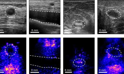
Three-dimensional transrectal ultrasonography (3D-TRUS) allows accurate 3D examination of pathological structures and prediction of extracapsular tumor extension.

Medisons Accuvix XQ Prestige 07 - a real-time 3D US scanner.
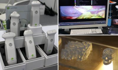
Medison's Accuvix XQ Prestige 07 - a real-time 3D US scanner.
180 randomly selected patiens with initial diagnosed prostate cancer participated in a study at the Medical University of Innsbruck. Dr. Michael Mitterberger and colleagues used 3D-TRUS, which provides ultrasonograms in three planes simultaneously, for local staging of prostate cancer.

“3D-TRUS can be used with no problems and is a simple procedure allowing exact assessement for staging prostate cancer,” said Dr, Mitterberger. “Locally confined and locally advanced prostate cancer can be differentiated with maximum convenience, reliability, and ease.”
3D-TRUS highly increase chances to detect pathological capsular perforation of the tumor: It predicted 44 of 53 cases correctly and features an overall accuracy of 96%.

The investigators conclude their report in the July issue of BJU International: “3D-Trus can be considered to guide the decision before a planned radical prostatectomy. If 3D-TRUS indicated locally advanced disease, the probability of capsular perforation or seminal vesicle invasion is very high.”

05.08.2007

More on the subject:

Related articles

Photo

News • Physics-driven framework

Quantum mechanics to remove noise from medical images

Medical imaging methods are often affected by background noise, which can obscure fine anatomical details. A new approach to solve this problem draws inspiration from quantum mechanics.

Photo

News • Ultrafast power doppler imaging

Monitoring brain blood flow during surgery to prevent stroke

A new method to monitor blood flow in the brain helps neurosurgeons detect the risk of a stroke during surgery – and potentially prevent it. This could also be useful for other types of operations.

Photo

News • Neural network screening tool

AI-enhanced ultrasound early detects cardiac amyloidosis

A neural network AI has been trained to detect cardiac amyloidosis from a single echocardiogram video of the heart's apical four-chamber view and differentiate it from similar heart conditions.

Related products

Subscribe to Newsletter