Image source: HUN-REN; credit: András Kriston
News • Deep visual multi-omics profiling
Digital pathology AI to break down tumor cell populations
A cancerous tumor is not a single enemy. It is made up of many different cell populations that can behave in very different ways. Among them, even at the time of diagnosis, there may already be cells that months or years later could give rise to metastases or become resistant to treatment. The challenge is that these critical cell populations are extremely difficult to detect in time. Researchers in Szeged, working together with Swiss and Swedish partners, have achieved an important breakthrough in this field.
The Momentum Microscopic Image Analysis and Machine Learning Research Group, led by Péter Horváth, recently appointed State Secretary for Science, is working to address this challenge. Based at the HUN-REN Biological Research Centre, Szeged, the group develops artificial intelligence-based methods that connect microscopic tissue images with molecular information. Two recent publications in the international scientific journals EMBO Molecular Medicine and npj Precision Oncology report, for the first time, single-cell-level measurements of the genetic and protein profiles of tumor clones, as well as the genetics of metastasis formation by tumor clones. These results open new perspectives for the future of precision cancer therapy.
This may bring us closer to understanding which cell populations can be responsible for the formation of metastases
Ede Migh
Traditional molecular analyses often examine an entire tissue sample at once. As a result, important differences between distinct regions of a tumor may be lost. The aim of the new approach is to enable researchers to see not only what a tumor looks like, but also how its different cell populations function. In this way, the internal map of a tumor becomes not only visible, but also readable at the molecular level.
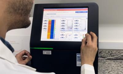
The group at the HUN-REN Biological Research Centre, Szeged develops these digital pathology and spatial omics technologies together with Swedish and Swiss partners. Key collaborators include Holger Moch, a molecular pathologist at University Hospital Zurich, and György Marko-Varga, research professor at Lund University. A major technological foundation of the work is the automated single-cell research center established by the Szeged group. This globally unique system enables AI-selected cells to be isolated with high precision, without human intervention, even around the clock, for subsequent molecular analysis.
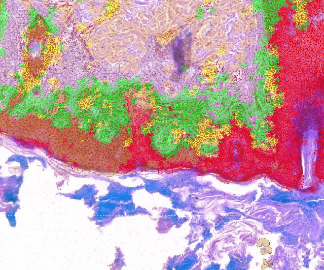
The approach is based on Deep Visual Proteomics, or DVP. In this method, artificial intelligence first analyzes histological tissue images and identifies relevant cell types or cell populations. Researchers then use a thin laser beam to precisely isolate these cells from the sample. Next, proteomic analysis is used to map which proteins are present in the selected cells. Proteins are particularly informative because they directly reflect how a cancer cell functions, grows, adapts, or becomes resistant to treatment.
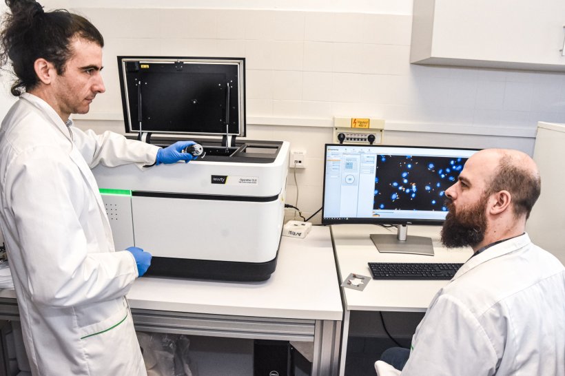
Image source: HUN-REN; credit: Petra Szili
In this study, the research group further developed DVP in an important way: the tumor cell populations selected by AI-based image analysis were examined not only by proteomics, meaning at the protein level, but also by transcriptomics. Transcriptomics shows which genes are active in a cell. This was particularly important for cell populations that appeared more dangerous under the microscope. The researchers were able to examine the same cell groups from two complementary perspectives: which genes were switched on in them, and what protein-level functions resulted from this activity.
With this approach, researchers can more precisely reveal how visually distinct cell populations within a tumor differ from each other biologically. This matters because the aggressiveness of a cancer cell is not explained by a single factor, but by the combined effects of gene activity, protein function, metabolism, and interactions with the immune system.
In another study, the research group demonstrated the clinical potential of the technology through the case of a young patient with recurrent metastatic melanoma. Samples from the patient’s original tumor, as well as later lung and brain metastases, were analyzed using AI-based digital pathology and spatially resolved proteomics.
Artificial intelligence identified two distinct tumor cell populations in the patient’s original melanoma. Cells in the later metastases mainly resembled one of these early populations. Protein-level analysis confirmed this observation: the molecular pattern of this cell population was closest to that of the metastatic lesions. This suggests that traces of the disease’s later spread may already have been present in the original tumor.
“In the melanoma case, we did not simply see that different parts of the tumor looked different. With this method, we could also show that these differences were reflected at the protein level. This may bring us closer to understanding which cell populations can be responsible for the formation of metastases,” said Ede Migh, biologist and one of the researchers involved in the study.

Image source: HUN-REN
The interconnected work of the Szeged research group shows that tumors can no longer be understood simply as uniform masses. Different cell populations can coexist within the same tumor, and some of them may play a crucial role in metastasis, aggressive growth, or resistance to treatment. In this approach, AI does not diagnose on its own. Instead, it provides a new level of resolution for studying tumors. While traditional pathological evaluation classifies larger tissue regions, artificial intelligence can analyze the sample cell by cell. This may reveal what types of cancer cells are present in different regions of the tumor, where they are located, what patterns they form, and in what proportions they occur.
“The more precisely we can see which cells drive the disease forward, the closer we get to targeting not only the tumor in general, but its most dangerous parts,” said Péter Horváth.
Although these findings do not yet represent an immediate new therapy available to all patients, they may help researchers and clinicians map tumor behavior more precisely in the future, better assess risks, and support more personalized treatment decisions.
Source: HUN-REN Szegedi Biológiai Kutatóközpont
13.08.2026