Article • Smarter predictions
Artificial Intelligence helping to detect breast cancer
Scientists are using Artificial Intelligence (AI) to support more effective breast cancer detection.
Report: Mark Nicholls
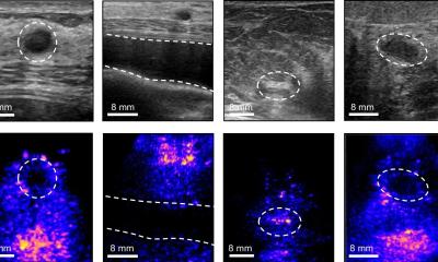
The researchers at Massachusetts Institute of Technology (MIT) Computer Science and Artificial Intelligence Laboratory (CSAIL), Massachusetts General Hospital (MGH), and Harvard Medical School, are using the machine learning system to predict whether breast lesions identified from a biopsy will turn out to be cancerous. Now, it is hoped the research could help reduce the number of unnecessary breast cancer surgeries because it is able to pinpoint which lesions are cancerous more accurately and more efficiently. In the study, the system was trained on information about such lesions and looked for patterns among a range of data points, including demographics, family history, biopsies and pathology reports.
Beyond a 'one-size-fits-all approach'
Because diagnostic tools are so inexact, there is an understandable tendency for doctors to over-screen for breast cancer
Regina Barzilay
When tested on 335 high-risk lesions, it correctly diagnosed 97% as malignant. The research team suggest that such levels of accuracy could lead to a reduction in the number of unnecessary surgeries by more than 30%. While mammograms can detect cancers, there is also a risk of false positive results that can lead to unnecessary biopsies and surgeries – often from “high-risk” lesions that appear suspicious on mammograms and have abnormal cells when tested by needle biopsy. The researchers say patients have the lesion surgically removed but it often turns out to be benign, meaning women going through unnecessary operations. To try to address this, the team developed the machine learning system to predict if a high-risk lesion identified on needle biopsy after a mammogram will upgrade to cancer at surgery.
Dr Regina Barzilay, MIT’s Delta Electronics Professor of Electrical Engineering and Computer Science, said: “Because diagnostic tools are so inexact, there is an understandable tendency for doctors to over-screen for breast cancer. When there’s this much uncertainty in data, machine learning is exactly the tool that we need to improve detection and prevent over-treatment. “A model like this will work anytime you have lots of different factors that correlate with a specific outcome. It hopefully will enable us to start to go beyond a one-size-fits-all approach to medical diagnosis.”
Into the random forest
Now we can have a more informed discussion with our patient about her options
Constance Lehman
Using a method known as a “random-forest classifier,” the model resulted in fewer unnecessary surgeries compared to the strategy of always doing surgery, while also being able to diagnose more cancerous lesions than the strategy of only doing surgery on traditional “high-risk lesions.” Dr Constance Lehman, Professor at Harvard Medical School and chief of the Breast Imaging Division at MGH’s Department of Radiology said: “To our knowledge, this is the first study to apply machine learning to the task of distinguishing high-risk lesions that need surgery from those that don’t. We believe this could support women to make more informed decisions about their treatment, and that we could provide more targeted approaches to health care in general.”
It is hoped that MGH radiologists will begin incorporating the model into their clinical practice over the next year. “In the past we might have recommended that all high-risk lesions be surgically excised,” Dr Lehman added. “But now, if the model determines that the lesion has a very low chance of being cancerous in a specific patient, we can have a more informed discussion with our patient about her options. It may be reasonable for some patients to have their lesions followed with imaging rather than surgically excised.”
The team – which also included Manisha Bahl, director of the Massachusetts General Hospital Breast Imaging Fellowship Program - says that they are working to further evolve the model and in the future hope to incorporate the actual images from the mammograms and images of the pathology slides, as well as more extensive patient information from medical records.
Profiles:
Dr Constance Lehman is a Professor at Harvard Medical School and chief of the Breast Imaging Division at MGH’s Department of Radiology. After graduating from Duke University and receiving medical and doctoral degrees at Yale University, she became professor and vice chair of Radiology and division chief of Breast Imaging at the Seattle Cancer Care Alliance before her recent move to MGH.
Dr Regina Barzilay is a Delta Electronics professor in the Department of Electrical Engineering and Computer Science and a member of the Computer Science and Artificial Intelligence Laboratory at the Massachusetts Institute of Technology. Her research interests are in natural language processing, applications of deep learning to chemistry and oncology.
Dr Manisha Bahl is a breast imaging radiologist and director of the Breast Imaging Fellowship Programme at Massachusetts General Hospital/Harvard Medical School in Boston. After graduating from the Harvard School of Public Health with an MPH in Health Policy and Management, she completed a radiology residency and breast imaging fellowship at Duke University Medical Centre and joined the faculty at Massachusetts General Hospital/Harvard Medical School in July 2016.
26.11.2017