Image source: Uppsala University; photo: Mikael Wallerstedt
News • Discovery of tumour indicating morphological changes
Prostate cancer: AI finds changes missed by pathologists
Using AI, researchers at Uppsala University have been able to find subtle tissue changes that allow prostate cancer to be detected long before it becomes visible to the human eye.
Using AI, researchers at Uppsala University have been able to find subtle tissue changes that allow the cancer to be detected long before it becomes visible to the human eye. The team published their findings in Scientific Reports.
Previous research has demonstrated that AI is able to detect tissue changes indicative of cancer. In the current study, the researchers show that AI can also find cancers missed by pathologists. “The study has been nicknamed the ‘missed study’, as the goal of finding the cancer was ‘missed’ by the pathologists. We have now shown that with the help of AI, it is possible to find signs of prostate cancer that were not observed by pathologists in more than 80% of samples from men who later developed cancer,” says Carolina Wählby, who led the AI development in the study.
This shows that AI analysis of routine biopsies can detect subtle signs indicating clinically significant prostate cancer before it becomes obvious to a pathologist
Carolina Wählby
The project is based on a collaboration with Umeå University, where the researchers collected samples from men called for sample-taking over a number of years. All 232 men in the study were assessed as healthy when their biopsies were examined by pathologists. After less than two-and-a-half years, half of the men in the study had developed aggressive prostate cancer, while the rest were still cancer-free eight years later.
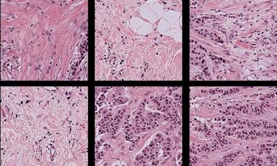
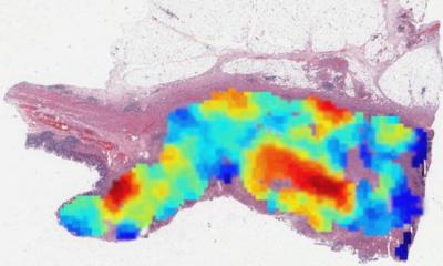
As all tissue samples were initially assessed as negative, the researchers developed a new way to train the AI tool. It was trained by analysing each biopsy image bit by bit, with the assumption that abnormal patterns ought to be present somewhere in the biopsies that came from patients who later developed aggressive cancer, while the other images should not contain such patterns. The AI was then tested on an independent set of images. “When we looked at the patterns that the AI ranked as informative, we saw changes in the tissue surrounding the glands in the prostate – changes also observed in other studies. This shows that AI analysis of routine biopsies can detect subtle signs indicating clinically significant prostate cancer before it becomes obvious to a pathologist,” says Wählby.
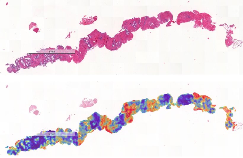
Image credit: Uppsala University/Carolina Wählby
The researchers suggest that this type of analysis could be used to decide how soon men who have been assessed as healthy should be followed up. The imaging data collected and the researchers’ methods are openly available for further research and development.
Source: Uppsala University
26.08.2025