Source: University of Waterloo
Article • Imaging
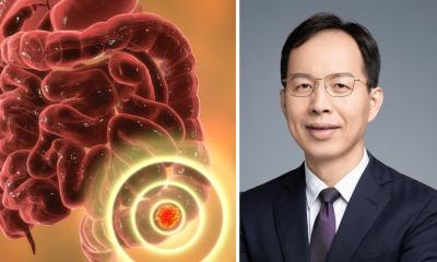
Photoacoustic technology could 'revolutionize' cancer surgery
Cancer treatment could be dramatically improved by an invention at the University of Waterloo to precisely locate the edges of tumors during surgery to remove them.
The new imaging technology uses the way light from lasers interacts with cancerous and healthy tissues to distinguish between them in real-time and with no physical contact, an advancement with the potential to eliminate the need for secondary surgeries to get missed malignant tissue. "This is the future, a huge step towards our ultimate goal of revolutionizing surgical oncology," said Parsin Haji Reza, a systems design engineering professor who leads the project. "Intraoperatively, during surgery, the surgeon will be able to see exactly what to cut and how much to cut."
Doctors now rely primarily on pre-operation MRI images and CT scans, experience and visual inspection to determine the margins of tumors during operations. Tissue samples are then sent to labs for testing, with waits of up to two weeks for results to show if the tumor was completely removed or not. In about 10 per cent of cases—the rates for different kinds of cancer involving tumors vary widely—some cancerous tissue has been missed and a second operation is required to remove it.
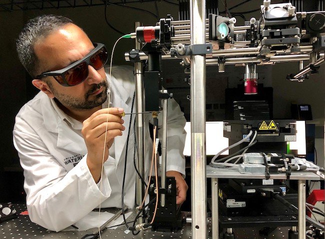
The photoacoustic technology developed at Waterloo works by sending laser light pulses into targeted tissue, which absorbs them, heats up, expands and produces soundwaves. A second laser reads those soundwaves, which are then processed to determine if the tissue is cancerous or non-cancerous.The system has already been used to make accurate images of even relatively thick, untreated human tissue samples for the first time ever, a key breakthrough in the development process.
Next steps include imaging fresh tissue samples taken during surgeries, integrating the technology into a surgical microscope and, finally, using the system directly on patients during operations. "This will have a tremendous impact on the economics of health-care, be amazing for patients and give clinicians a great new tool," said Haji Reza, director of the PhotoMedicine Labs at Waterloo. "It will save a great deal of time and money and anxiety."
Researchers hope to develop a fully functioning system within about two years, a process including the need to clear ethical hurdles and securing regulatory approvals.
Source: University of Waterloo
18.09.2019