© samunella – stock.adobe.com
Article • AI-powered prediction
Machine learning identifies cardiotoxicity risk in breast cancer patients
Researchers have developed a machine learning algorithm that uses cardiac MRI images to help identify breast cancer patients who may be at risk of cardiotoxicity during cancer treatment. The research, led by cardiologist Dr Paaladinesh Thavendiranathan from Toronto General Hospital University Health Network, was presented at the European Society of Cardiology's Cardio-Oncology Conference in Florence in June.
By Mark Nicholls

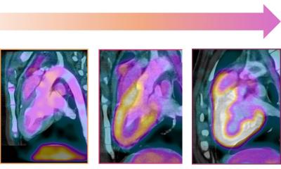
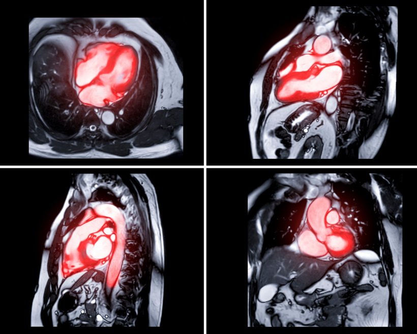
Image courtesy of Dr Thavendiranathan
The study addressed the challenge of predicting cancer therapy-related cardiac dysfunction (CTRCD), which the researchers noted "remains a challenge." According to the research paper, which was published in European Heart Journal Supplements, clinical risk models and conventional cardiac magnetic resonance (CMR) analysis ‘are limited’ in predicting HER2+ targeted therapy (HER2-TT) CTRCD risk.
Dr Thavendiranathan, who is also a Professor of Medicine at the University of Toronto, Canada, explained that current techniques for risk stratification ‘are not very effective.’
Promising study performance
The Canadian study aimed to determine if deep learning (DL) approaches using CMR cine images pre or early during cancer therapy can predict CTRCD better than clinical risk scores or conventional quantified imaging measures.
Women with early-stage HER2+ breast cancer receiving sequential anthracyclines and trastuzumab from three prospective studies were included. Patients were seen pre- and post-anthracycline and sequentially during treatment with repeated cardiac imaging (echocardiography and CMR) to create various machine learning models to predict CTRCD.
‘This model performed better than clinical risk factors, echocardiography measures, and traditional cardiac MRI measures or biomarkers,’ Dr Thavendiranathan said. ‘When we validated our findings in a separate cohort, the model continued to perform well.’
Recommended article

Article • Five-year EU project to avoid heart damage in oncology patients
Cardiac collaterals in breast cancer therapy
Modern cancer therapies are tough on the tumours, but often, also on the heart of the patients. The “CARDIOCARE” project aims to reduce the cardiac burden of anti-cancer therapies through more patient-tailored treatment approaches. At the ESC cardiology congress, Professor Katerina Naka from the project’s consortium explained why older patients are at the highest risk of cardiotoxic…
Using information from the whole heart
The research concluded that ‘in women with breast cancer receiving anthracyclines and HER2-TT, a DL model using CMR short axis cine images pre-anthracycline had higher discrimination for future CTRCD than clinical and conventional imaging quantification models.’
If patients at risk for cardiotoxicity can be identified even before cancer therapy, preventive strategies can be instituted to reduce the risk of cardiotoxicity
Paaladinesh Thavendiranathan
Dr Thavendiranathan's key finding was that a machine learning algorithm applied to a single set of cardiac MRI images could identify patients at high risk of cardiotoxicity. He suggested this ‘may be due to the ability to potentially use information from the whole heart with the ML algorithm to risk stratify patients, as opposed to single chamber information as we do traditionally.’
The expert outlined potential clinical benefits of identifying at-risk patients before treatment begins: ‘This would provide opportunities for clinicians to cater their cancer therapies to minimize risk or to add cardioprotective medications or to modify the surveillance strategies to pick up early cardiac dysfunction during cancer therapy,’ he said. ‘If patients at risk for cardiotoxicity can be identified even before cancer therapy, preventive strategies can be instituted to reduce the risk of cardiotoxicity. This will reduce the chance of the patient subsequently developing heart failure or having their life saving cancer therapy interrupted.’
Transforming risk stratification
The next step, according to Dr Thavendiranathan, is validation of findings in larger cohorts and to consider applying similar methods to more readily available imaging tests in patients with cancers, such as echocardiography. He expressed hope that further research could potentially lead to a baseline test for use prior to cancer therapy that may identify patients at risk of developing cardiotoxicity. ‘This has the potential to transform the way that we risk stratify patients as current techniques are not very effective and hopefully, by early treatment, have an impact on the cardiovascular prognosis and overall survival of these patients,’ Dr Thavendiranathan concluded.
The session at ESC Cardio-Oncology 2025 also included presentations on non-uniform cardiovascular risks of tyrosine kinase inhibitors, left atrial strain in multiple myeloma patients undergoing carfilzomib therapy, cardiovascular adverse events with cdk4/6 inhibitors, cardiac function following anti-BCMA CAR-T therapy, and cardiovascular safety of modern radiotherapy techniques in breast cancer.
Profile:
Dr Paaladinesh Thavendiranathan is a cardiologist at the Toronto General Hospital University Health Network, and a Professor of Medicine at the University of Toronto, Canada. With a background in advanced cardiovascular imaging and an international expert in the field of cardio-oncology, his research is in the use of advanced cardiac imaging techniques for detection and management of cardiac toxicity. He is Director of the Ted Rogers Program in Cardiotoxicity Prevention which focuses on cardiac toxicity from systemic therapies including cancer therapy.
01.09.2025